 Explore
 Validate
 Learn
Learn Western blot
 Flow cytometry
Antibody data
- Antibody Data
- Antigen structure
- References [0]
- Comments [0]
- Validations
- Western blot [1]
- Immunocytochemistry [1]
- Other assay [2]
Submit
Validation data
Reference
Comment
Report error
- Product number
- 702230 - Provider product page

- Provider
- Invitrogen Antibodies
- Product name
- Phospho-GSK3 alpha/beta (Tyr279, Tyr216) Recombinant Rabbit Monoclonal Antibody (19H1L12)
- Antibody type
- Monoclonal
- Antigen
- Synthetic peptide
- Reactivity
- Human, Mouse
- Host
- Rabbit
- Isotype
- IgG
- Antibody clone number
- 19H1L12
- Vial size
- 100 µg
- Concentration
- 0.5 mg/mL
- Storage
- Store at 4°C short term. For long term storage, store at -20°C, avoiding freeze/thaw cycles.
No comments: Submit comment
Supportive validation
- Submitted by
- Invitrogen Antibodies (provider)
- Main image

- Experimental details
- Western blot analysis was performed on Whole cell extracts (30 µg lysate) of 3T3-L1 (Lane 1), 3T3-L1 treated with Insulin (100nM-10min) (Lane 2), A549 (Lane 3) and A549 treated with Insulin (100nM-10min) (Lane 4). The blots were probed with Anti-GSK3a (pY279) + GSK3b (pY216) Recombinant Rabbit Polyclonal Antibody (Product # 702230, 1-2 µg/mL) and detected by chemiluminescence using Goat anti-Rabbit IgG (H+L) Superclonal Secondary Antibody, HRP conjugate (Product # A27036, 0.4 µg/mL, 1:2500 dilution). Two isoforms of 46 kDa and 51 kDa band corresponding to GSK3a (pY279)+GSK3b (pY216) was observed. To confirm the specificity of Antibody, competition was performed with the phosphopeptide (10 µg/mL) as shown in the corresponding blot on the right. The phosphopeptide competes with the antibody and prevents it from binding to the target protein. Known quantity of protein samples were electrophoresed using Novex® NuPAGE® 4-12% Bis-Tris gel (Product # NP0321BOX), XCell SureLock Electrophoresis System (Product # EI0002) and Novex® Sharp Pre-Stained Protein Standard (Product # LC5800). Resolved proteins were then transferred onto a nitrocellulose membrane with iBlot® Dry Blotting System (Product # IB21001). The membrane was probed with the relevant primary and secondary Antibody following blocking with 5% skimmed milk. Chemiluminescent detection was performed using Pierce™ ECL Western blotting Substrate (Product # 32106).

Supportive validation
- Submitted by
- Invitrogen Antibodies (provider)
- Main image
- Experimental details
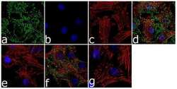
- For immunofluorescence analysis, 3T3-L1 cells were fixed and permeabilized for detection of endogenous CSL/RBP J using GSK-3 alpha (pY279) + GSK3 beta (pY216) Recombinant Rabbit Monoclonal Antibody (Product # 702230, 2 µg/mL) and labeled with Goat anti-Rabbit IgG (H+L) Superclonal Secondary Antibody, Alexa Fluor® 488 conjugate (Product # A27034, 1:2000). Panel a) shows representative cells that were stained for detection and localization of GSK-3 alpha (pY279) + GSK3 beta (pY216) protein (green), Panel b) is stained for nuclei (blue) using SlowFade® Gold Antifade Mountant with DAPI (Product # S36938). Panel c) represents cytoskeletal F-actin staining using Alexa Fluor® 555 Rhodamine Phalloidin (Product # R415, 1:300). Panel d) is a composite image of Panels a, b and c clearly demonstrating nuclear and cytoplasmic localization of GSK-3 alpha (pY279) + GSK3 beta (pY216). Antibody specificity was demonstrated by competition with the GSK-3 alpha (pY279) + GSK3 beta (pY216) phospho peptide, which results in loss of signal in panel e). Panel f) shows no competition with the non-phospho peptide. The images were captured at 60X magnification

Supportive validation
- Submitted by
- Invitrogen Antibodies (provider)
- Main image

- Experimental details
- Figure 6 Fap1-inhibition with SLV peptide increases phosphorylation of Fap1-substrates Fas and Gsk3beta in a murine xenograft model SW620 cells were injected in the flanks of athymic Nude mice and tumor volume was determination biweekly. Mice were treated weekly with oxaliplatin (days 0, 7 and 14) and injected daily with Fap1 blocking SLV peptide or VLS control peptide, or treated with SLV or VLS peptide alone (n=12 per cohort). Tumors were simultaneously harvested from cohorts of mice when control tumors were >2,000 mm3. (A) SLV peptide increases gland formation in xenograft tumors with or without oxaliplatin. Histology was analyzed by hematoxylin/ eosin staining. Fap1 expression was determined by immunofluorescence. Relative fluorescent intensity (RFI) of Fap1 staining is indicated below relevant panels. (B) SLV peptide increases Fas-phosphorylation in xenograft tumors with or without by oxaliplatin. Immunofluorescent detection of total versus phospho-Fas was performed with DAPI staining of nuclei. Areas without gland formation were selected for this study. (C) SLV peptide increases Gsk3beta-phosphorylation with or without oxaliplatin. Immunofluorescent detection of total versus phospho- Gsk3beta was performed with DAPI staining of nuclei. Areas without gland formation were selected for this study.

- Submitted by
- Invitrogen Antibodies (provider)
- Main image

- Experimental details
- Figure 7 Fap1-inhibition with SLV peptide increases Fas and Gsk3beta phosphorylation in CD133 + cells in a murine xenograft model SW620 cells were injected in the flanks of athymic Nude mice and tumor volume was determination biweekly. Mice were treated weekly with oxaliplatin (days 0, 7 and 14) and injected daily with Fap1 blocking SLV peptide or VLS control peptide, or treated with SLV or VLS peptide alone (n=12 per cohort). Tumors were simultaneously harvested from cohorts of mice when control tumors were >2,000 mm3. (A) SLV peptide increases Fas phosphorylation in CD133 + xenograft tumors with or without oxaliplatin. Immunofluorescent detection of phospho-Fas or CD133 was performed with DAPI staining of nuclei. (B) SLV peptide increases Gsk3beta phosphorylation in CD133 + xenograft tumors with or without oxaliplatin. Immunofluorescent detection of phospho-Gsk3beta or CD133 was performed with DAPI staining of nuclei.
