Explore
 Validate
 Learn
Learn Western blot
 Immunocytochemistry
 Immunohistochemistry
Antibody data
- Antibody Data
- Antigen structure
- References [6]
- Comments [0]
- Validations
- Immunocytochemistry [2]
- Other assay [1]
Submit
Validation data
Reference
Comment
Report error
- Product number
- MA5-14873 - Provider product page

- Provider
- Invitrogen Antibodies
- Product name
- Phospho-GSK3B (Ser9) Monoclonal Antibody (C.367.3)
- Antibody type
- Monoclonal
- Antigen
- Synthetic peptide
- Description
- It is not recommended to aliquot this antibody.
- Reactivity
- Human, Mouse, Rat
- Host
- Rabbit
- Isotype
- IgG
- Antibody clone number
- C.367.3
- Vial size
- 100 μL
- Concentration
- 57 μg/mL
- Storage
- -20°C
Submitted references The neuroprotective mechanism of lithium after ischaemic stroke.
MYBL2 in synergy with CDC20 promotes the proliferation and inhibits apoptosis of gastric cancer cells.
The extracellular HDAC6 ZnF UBP domain modulates the actin network and post-translational modifications of Tau.
Sevoflurane inhibits the proliferation and invasion of hepatocellular carcinoma cells through regulating the PTEN/Akt/GSK‑3β/β‑catenin signaling pathway by downregulating miR‑25‑3p.
ATM-associated signalling triggers the unfolded protein response and cell death in response to stress.
Lithium treatment decreases activities of tau kinases in a murine model of senescence.
Chen B, Zhang M, Ji M, Zhang D, Chen B, Gong W, Li X, Zhou Y, Dong C, Wen G, Zhan X, Wu X, Yuan H, Xu E, Xia M, Verkhratsky A, Li B
Communications biology 2022 Feb 3;5(1):105
Communications biology 2022 Feb 3;5(1):105
MYBL2 in synergy with CDC20 promotes the proliferation and inhibits apoptosis of gastric cancer cells.
Deng Q, Wu L, Li Y, Zou L
Advances in clinical and experimental medicine : official organ Wroclaw Medical University 2021 Sep;30(9):957-966
Advances in clinical and experimental medicine : official organ Wroclaw Medical University 2021 Sep;30(9):957-966
The extracellular HDAC6 ZnF UBP domain modulates the actin network and post-translational modifications of Tau.
Balmik AA, Sonawane SK, Chinnathambi S
Cell communication and signaling : CCS 2021 May 1;19(1):49
Cell communication and signaling : CCS 2021 May 1;19(1):49
Sevoflurane inhibits the proliferation and invasion of hepatocellular carcinoma cells through regulating the PTEN/Akt/GSK‑3β/β‑catenin signaling pathway by downregulating miR‑25‑3p.
Cao Y, Lv W, Ding W, Li J
International journal of molecular medicine 2020 Jul;46(1):97-106
International journal of molecular medicine 2020 Jul;46(1):97-106
ATM-associated signalling triggers the unfolded protein response and cell death in response to stress.
Hotokezaka Y, Katayama I, Nakamura T
Communications biology 2020 Jul 14;3(1):378
Communications biology 2020 Jul 14;3(1):378
Lithium treatment decreases activities of tau kinases in a murine model of senescence.
Tajes M, Gutierrez-Cuesta J, Folch J, Ferrer I, Caballero B, Smith MA, Casadesus G, Camins A, Pallás M
Journal of neuropathology and experimental neurology 2008 Jun;67(6):612-23
Journal of neuropathology and experimental neurology 2008 Jun;67(6):612-23
No comments: Submit comment
Supportive validation
- Submitted by
- Invitrogen Antibodies (provider)
- Main image

- Experimental details
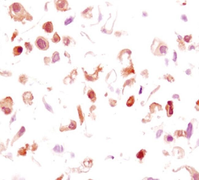
- Immunocytochemical analysis of Phospho-GSK-3beta pSer9 in paraffin-embedded wild type (left) or GSK-3beta (-/-) MEFs using a Phospho-GSK-3beta pSer9 monoclonal antibody (Product # MA5-14873).

- Submitted by
- Invitrogen Antibodies (provider)
- Main image

- Experimental details
- Immunocytochemical analysis of Phospho-GSK-3beta pSer9 in paraffin-embedded wild type (left) or GSK-3beta (-/-) MEFs using a Phospho-GSK-3beta pSer9 monoclonal antibody (Product # MA5-14873).
Supportive validation
- Submitted by
- Invitrogen Antibodies (provider)
- Main image
- Experimental details
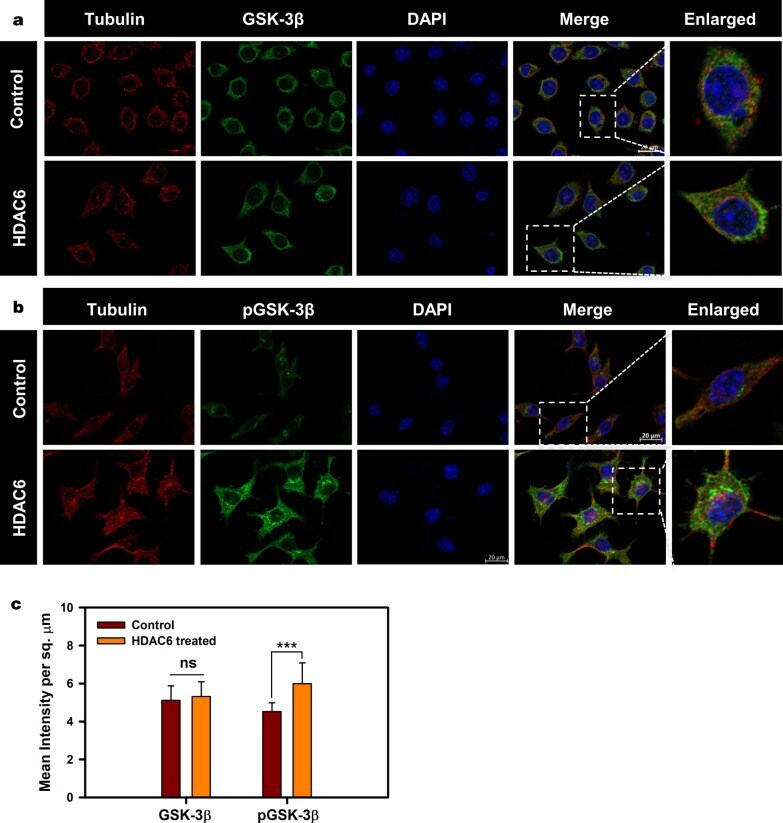
- Fig. 1 Downregulation of GSK-3beta activity by HDAC6. a Neuro2a mapped for total GSK-3beta shows their unaltered levels upon HDAC6 ZnF UBP. b Inhibitory phosphorylation of GSK-3beta at Ser9 increases upon HDAC6 ZnF UBP treatment. The enlarged image shows the elevated level of pGSK-3beta compared to neuro2a cell control. c Quantification of mean fluorescence intensity for GSK-3beta in cell control and HDAC6 ZnF UBP treated cells showed non-significant difference while the levels of pGSK-3beta were significantly increased upon HDAC6 ZnF UBP treatment in comparison to cell control