Explore
 Validate
 Learn
Learn Western blot
Antibody data
- Antibody Data
- Antigen structure
- References [3]
- Comments [0]
- Validations
- Western blot [1]
Submit
Validation data
Reference
Comment
Report error
- Product number
- OPA1-03083 - Provider product page

- Provider
- Invitrogen Antibodies
- Product name
- Anti-Phospho-GSK3 alpha/beta (Tyr279, Tyr216) Polyclonal Antibody
- Antibody type
- Polyclonal
- Antigen
- Synthetic peptide
- Description
- OPA1-03083 detects the alpha and beta isoforms of phospho-GSK3 from human and mouse tissues. This antibody is expected to cross-react with rat, frog, and zebra fish GSK-3 due to 100% sequence homology. OPA1-03083 has been successfully used in Western blot procedures. By Western blot, this antibody detects an ~51 kDa protein and an ~47 kDa protein from stimulated 3T3-L1 cells representing tyrosine 279 phosphorylated GSK3alpha and tyrosine 216 phosphorylated GSK3beta, respectively. Positive control: serum starved mouse 3T3-L1 cells +/- insulin (100 nM, 10 minutes), or background extracts +/- human recombinant GSK-3â. OPA1-03083 immunizing phosphopeptide was derived from a regions of human GSK3alpha/beta that contain tyrosine 279/216.
- Reactivity
- Human, Mouse
- Host
- Rabbit
- Isotype
- IgG
- Vial size
- 100 µL
- Storage
- -20° C, Avoid Freeze/Thaw Cycles
Submitted references Silver nanoparticles induce neurotoxicity in a human embryonic stem cell-derived neuron and astrocyte network.
Exploiting an Asp-Glu "switch" in glycogen synthase kinase 3 to design paralog-selective inhibitors for use in acute myeloid leukemia.
The intersection of genetic and chemical genomic screens identifies GSK-3α as a target in human acute myeloid leukemia.
Repar N, Li H, Aguilar JS, Li QQ, Drobne D, Hong Y
Nanotoxicology 2018 Mar;12(2):104-116
Nanotoxicology 2018 Mar;12(2):104-116
Exploiting an Asp-Glu "switch" in glycogen synthase kinase 3 to design paralog-selective inhibitors for use in acute myeloid leukemia.
Wagner FF, Benajiba L, Campbell AJ, Weïwer M, Sacher JR, Gale JP, Ross L, Puissant A, Alexe G, Conway A, Back M, Pikman Y, Galinsky I, DeAngelo DJ, Stone RM, Kaya T, Shi X, Robers MB, Machleidt T, Wilkinson J, Hermine O, Kung A, Stein AJ, Lakshminarasimhan D, Hemann MT, Scolnick E, Zhang YL, Pan JQ, Stegmaier K, Holson EB
Science translational medicine 2018 Mar 7;10(431)
Science translational medicine 2018 Mar 7;10(431)
The intersection of genetic and chemical genomic screens identifies GSK-3α as a target in human acute myeloid leukemia.
Banerji V, Frumm SM, Ross KN, Li LS, Schinzel AC, Hahn CK, Kakoza RM, Chow KT, Ross L, Alexe G, Tolliday N, Inguilizian H, Galinsky I, Stone RM, DeAngelo DJ, Roti G, Aster JC, Hahn WC, Kung AL, Stegmaier K
The Journal of clinical investigation 2012 Mar;122(3):935-47
The Journal of clinical investigation 2012 Mar;122(3):935-47
No comments: Submit comment
Supportive validation
- Submitted by
- Invitrogen Antibodies (provider)
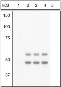
- Main image
- Experimental details
- Up-regulation and Antibody-Peptide Competition: Peptide Competition. Extracts of 3T3L1 cells stimulated with 100 nM insulin for 10 minutes were resolved by SDS-PAGE on a 10% Tris-glycine gel and transferred to PVDF. The membrane was blocked with a 5% BSA-TBST buffer for one hour at room temperature and either left untreated (1-4) or treated with lambda phosphatase (5), and then incubated with the GSK-3a (pY279)/beta (pY216) antibody (Product # OPA1-03083) for two hours at room temperature in a 1% BSA-TBST buffer, following its prior incubation with: the phosphopeptide immunogen (1), no peptide (2), the non-phosphopeptide corresponding to the phosphopeptide immunogen (3), or a generic phosphotyrosine-containing peptide (4). After washing, the membrane was incubated with goat F (ab')2 anti-rabbit IgG HRP conjugate (Product # ALI4404), and signals were detected using the Pierce SuperSignal™ method. The data show that only the phosphopeptide corresponding to GSK-3a (pY279)/beta (pY216) blocks the antibody signal, demonstrating the specificity of the antibody. The data also show that phosphatase stripping eliminates the signal, further verifying that the antibody is phospho-specific.