Explore
 Validate
 Learn
Learn Immunocytochemistry
Antibody data
- Antibody Data
- Antigen structure
- References [3]
- Comments [0]
- Validations
- Immunocytochemistry [1]
- Immunohistochemistry [1]
- Flow cytometry [1]
- Other assay [3]
Submit
Validation data
Reference
Comment
Report error
- Product number
- MA5-13122 - Provider product page

- Provider
- Invitrogen Antibodies
- Product name
- CD35 Monoclonal Antibody (E11)
- Antibody type
- Monoclonal
- Antigen
- Other
- Description
- MA5-13122 targets CD35 in FACS, ICC/IF, and IHC (P) applications and shows reactivity with Human and Non-human primate samples. The MA5-13122 immunogen is intact human monocytes.
- Reactivity
- Human
- Host
- Mouse
- Isotype
- IgG
- Antibody clone number
- E11
- Vial size
- 500 µL
- Concentration
- 0.8 mg/mL
- Storage
- 4° C
Submitted references Lymphoid Aggregates in the CNS of Progressive Multiple Sclerosis Patients Lack Regulatory T Cells.
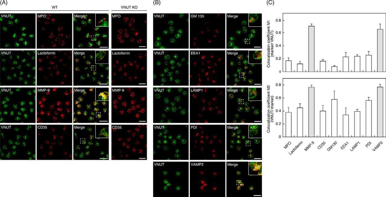
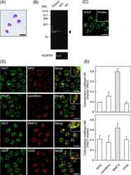
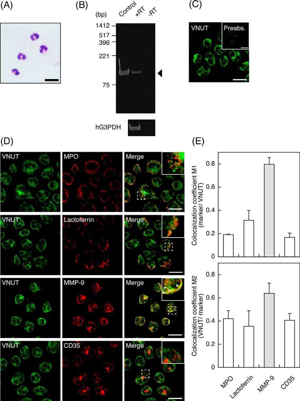
Vesicular nucleotide transporter mediates ATP release and migration in neutrophils.
Effect of B-cell depletion on coreceptor switching in R5 simian-human immunodeficiency virus infection of rhesus macaques.
Bell L, Lenhart A, Rosenwald A, Monoranu CM, Berberich-Siebelt F
Frontiers in immunology 2019;10:3090
Frontiers in immunology 2019;10:3090
Vesicular nucleotide transporter mediates ATP release and migration in neutrophils.
Harada Y, Kato Y, Miyaji T, Omote H, Moriyama Y, Hiasa M
The Journal of biological chemistry 2018 Mar 9;293(10):3770-3779
The Journal of biological chemistry 2018 Mar 9;293(10):3770-3779
Effect of B-cell depletion on coreceptor switching in R5 simian-human immunodeficiency virus infection of rhesus macaques.
Tasca S, Zhuang K, Gettie A, Knight H, Blanchard J, Westmoreland S, Cheng-Mayer C
Journal of virology 2011 Apr;85(7):3086-94
Journal of virology 2011 Apr;85(7):3086-94
No comments: Submit comment
Supportive validation
- Submitted by
- Invitrogen Antibodies (provider)
- Main image

- Experimental details
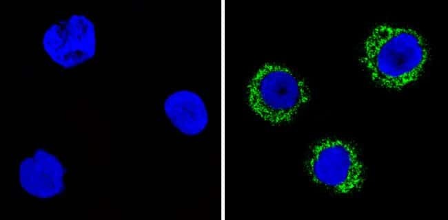
- Immunofluorescent analysis of CD35 (green) showing staining in the cytoplasm and membrane of THP-1 cells (right) compared to a negative control without primary antibody (left). Formalin-fixed cells were permeabilized with 0.1% Triton X-100 in TBS for 5-10 minutes and blocked with 3% BSA-PBS for 30 minutes at room temperature. Cells were probed with a CD35 monoclonal antibody (Product # MA5-13122) in 3% BSA-PBS at a dilution of 1:20 and incubated overnight at 4 ºC in a humidified chamber. Cells were washed with PBST and incubated with a DyLight-conjugated secondary antibody in PBS at room temperature in the dark. F-actin (red) was stained with a fluorescent red phalloidin and nuclei (blue) were stained with Hoechst or DAPI. Images were taken at a magnification of 60x.
Supportive validation
- Submitted by
- Invitrogen Antibodies (provider)
- Main image
- Experimental details
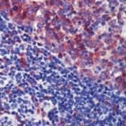
- Formalin-fixed, paraffin-embedded human tonsil stained with CD35 antibody using peroxidase-conjugate and AEC chromogen. Note cell membrane staining of follicular dendritic cells, B cells and granulocytes.

Supportive validation
- Submitted by
- Invitrogen Antibodies (provider)
- Main image
- Experimental details
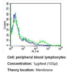
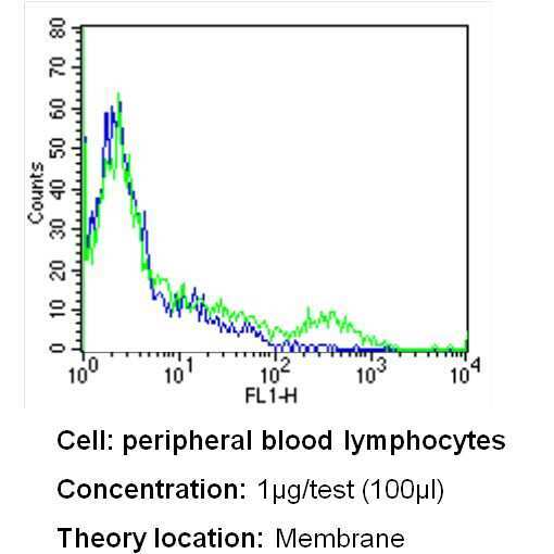
- Flow cytometry analysis of CD35 in PBMC cells (green) compared to an isotype control (blue). Human blood was collected, combined with a hydrophilic polysaccharide, centrifuged, transferred to a conical tube and washed with PBS. 50 µL of cell solution was added to each tube at a dilution of 2x10^7 cells/mL, followed by the addition of 50 µL of isotype control and primary antibody (Product # MA5-13122) at a dilution of 1 µg/test. Cells were incubated for 30 min at 4ºC and washed with a cell buffer, followed by incubation with a DyLight 488-conjugated secondary antibody for 30 min at 4ºC in the dark. FACS analysis was performed using 400 µL of cell buffer.
Supportive validation
- Submitted by
- Invitrogen Antibodies (provider)
- Main image
- Experimental details
- NULL
- Submitted by
- Invitrogen Antibodies (provider)
- Main image
- Experimental details
- NULL
- Submitted by
- Invitrogen Antibodies (provider)
- Main image
- Experimental details
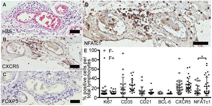
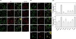
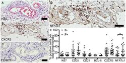
- Figure 3 Follicle-like structures of SPMS brains are devoid of FOXP3 expression, but exhibit NFATc1 + cells. (A) Representative meningeal follicle-like structure of SPMS spinal cord, which was screened based on H&E staining and characterized by >60 lymphocytes (score 3), detection of Ki67 + , CD35 + /CD21 + , and CD138 + cells on serial sections, termed F+. F-, if no or not all criteria were fulfilled. (B) Follicle-like structures could be characterized by CXCR5 + , (C) but not by FOXP3 + cells. (D) NFATc1 + cells were present in follicle-like structures. (E) IHC stainings of Ki67, CD35, CD21, BCL-6, CXCR5, and NFATc1 were quantified as frequency of total cells in follicle-like structures (F+) and less defined infiltrates (F-). Ki67 : F-, M = 1.52, SD = 3.73, n = 45; F+, M = 1.89, SD = 3.55, n = 38; Mann Whitney test, U = 734.0, p = 0.161. CD35 : F-, M = 15.41, SD = 22.95, n = 22; F+, M = 19.05, SD = 14.75, n = 21; Mann Whitney test, U = 159.0, p = 0.081. CD21 : F-, M = 5.68, SD = 10.63, n = 21; F+, M = 3.51, SD = 5.60, n = 20; Mann Whitney test, U = 183.5, p = 0.488. BCL-6 : F-, M = 0.32, SD = 1.08, n = 44; F+, M = 0.49, SD = 1.62, n = 38; Mann Whitney test, U = 803.0, p = 0.642. CXCR5 : F-, M = 14.72, SD = 17.24, n = 30; F+, M = 20.11, SD = 15.73, n = 29; Mann Whitney test, U = 319.0, p = 0.080. NFATc1 : F-, M = 7.15, SD = 11.68, n = 28; F+, M = 16.36, SD = 21.46, n = 31; Mann Whitney test, U = 307.0, p = 0.043. Scale bars A-D indicate 100 mum. *, p < 0.05.