Explore
 Validate
 Learn
Learn Western blot
 Immunocytochemistry
Antibody data
- Antibody Data
- Antigen structure
- References [4]
- Comments [0]
- Validations
- Western blot [1]
Submit
Validation data
Reference
Comment
Report error
- Product number
- GTX25813 - Provider product page

- Provider
- GeneTex
- Proper citation
- GeneTex Cat#GTX25813, RRID:AB_384130
- Product name
- PKC iota / lambda (phospho Thr563) antibody
- Antibody type
- Polyclonal
- Reactivity
- Human, Mouse, Canine
- Host
- Rabbit
Submitted references Conditional knockout of polarity complex (atypical) PKCι reveals an anti-inflammatory function mediated by NF-κB.
Myosin 5b loss of function leads to defects in polarized signaling: implication for microvillus inclusion disease pathogenesis and treatment.
The BAG-1 isoform BAG-1M regulates keratin-associated Hsp70 chaperoning of aPKC in intestinal cells during activation of inflammatory signaling.
Par-complex aPKC and Par3 cross-talk with innate immunity NF-κB pathway in epithelial cells.
Forteza R, Figueroa Y, Mashukova A, Dulam V, Salas PJ
Molecular biology of the cell 2016 Jul 15;27(14):2186-97
Molecular biology of the cell 2016 Jul 15;27(14):2186-97
Myosin 5b loss of function leads to defects in polarized signaling: implication for microvillus inclusion disease pathogenesis and treatment.
Kravtsov D, Mashukova A, Forteza R, Rodriguez MM, Ameen NA, Salas PJ
American journal of physiology. Gastrointestinal and liver physiology 2014 Nov 15;307(10):G992-G1001
American journal of physiology. Gastrointestinal and liver physiology 2014 Nov 15;307(10):G992-G1001
The BAG-1 isoform BAG-1M regulates keratin-associated Hsp70 chaperoning of aPKC in intestinal cells during activation of inflammatory signaling.
Mashukova A, Kozhekbaeva Z, Forteza R, Dulam V, Figueroa Y, Warren R, Salas PJ
Journal of cell science 2014 Aug 15;127(Pt 16):3568-77
Journal of cell science 2014 Aug 15;127(Pt 16):3568-77
Par-complex aPKC and Par3 cross-talk with innate immunity NF-κB pathway in epithelial cells.
Forteza R, Wald FA, Mashukova A, Kozhekbaeva Z, Salas PJ
Biology open 2013;2(11):1264-9
Biology open 2013;2(11):1264-9
No comments: Submit comment
Supportive validation
- Submitted by
- GeneTex (provider)
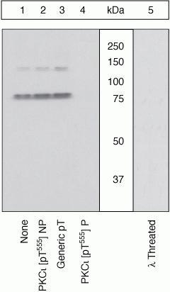
- Main image
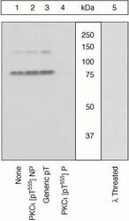
- Experimental details
- Western blot using PKC-iota [pT555]/PKC Lambda [pT563] Polyclonal Antibody, Rabbit (GTX25813). Lysates prepared from Jurkat cells stimulated with PMA were resolved by SDS-PAGE on a 10% polyacrylamide gel and transferred to PVDF. Membranes were either left untreated (1-4) or treated with lambda phosphatase (5), blocked with a 5% low-fat milk-TBST buffer for one hour at room temperature, and incubated with PKCiota [pT555] antibody for two hours at room temperature in a 3% low-fat milk-TBST buffer, following prior incubation with: no peptide (1), the non-phosphopeptide corresponding to the immunogen (2), a generic phosphothreonine-containing peptide (3), or, the phosphopeptide immunogen (4). After washing, membranes were incubated with goat F(ab¡¦)2 anti-rabbit IgG HRP conjugate and bands were detected. The data show that the phosphopeptide corresponding to PKCiota [pT555] blocks the antibody signal. The peptide corresponding to PKCzeta [pT560] blocks the antibody signal and the peptides corresponding to PKC isoforms betaI [pT642] and gamma [pT655] partially block the antibody signal (data not shown), suggesting cross-reactivity of the antibody with these sites. The antibody signal was not blocked by the corresponding peptides of any other PKC isoforms. The data also show that phosphatase stripping eliminates the signal, verifying that the antibody is phospho-specific