 Explore
 Validate
 Learn
Learn Western blot
 Immunocytochemistry
Antibody data
- Antibody Data
- Antigen structure
- References [0]
- Comments [0]
- Validations
- Immunocytochemistry [3]
- Flow cytometry [2]
Submit
Validation data
Reference
Comment
Report error
- Product number
- MA1-10449 - Provider product page

- Provider
- Invitrogen Antibodies
- Product name
- SHIP1 Monoclonal Antibody (SHIP-01)
- Antibody type
- Monoclonal
- Antigen
- Synthetic peptide
- Description
- This antibody reacts with SHIP-1, a phosphoinositide phosphatase largely confined to hematopoietic cells (intracellular antigen). Multiple forms of SHIP-1 have been reported with molecular weights of 110, 125, 130, 135 and 145 kDa. Western Blot: Reducing conditions.
- Reactivity
- Human
- Host
- Mouse
- Isotype
- IgG
- Antibody clone number
- SHIP-01
- Vial size
- 100 μg
- Concentration
- 1 mg/mL
- Storage
- 4°C, do not freeze
No comments: Submit comment
Supportive validation
- Submitted by
- Invitrogen Antibodies (provider)
- Main image
- Experimental details
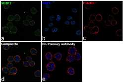
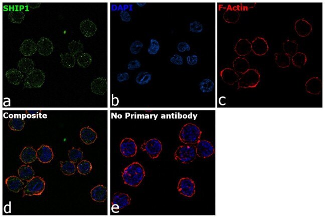
- Immunofluorescence analysis of SHIP1 was performed using 70% confluent log phase THP-1 cells. The cells were fixed with 4% paraformaldehyde for 10 minutes, permeabilized with 0.1% Triton™ X-100 for 15 minutes, and blocked with 2% BSA for 1 hour at room temperature. The cells were labeled with SHIP1 Mouse Monoclonal Antibody (SHIP-01) (Product # MA1-10449) at 1:100 dilution in 0.1% BSA, incubated at 4 degree Celsius overnight and then with Goat anti-Mouse IgG (H+L) Highly Cross-Adsorbed Secondary Antibody, Alexa Fluor Plus 488 (Product # A32723) at a dilution of 1:2000 for 45 minutes at room temperature (Panel a: green). Nuclei (Panel b: blue) were stained with ProLong™ Diamond Antifade Mountant with DAPI (Product # P36962). F-actin (Panel c: red) was stained with Rhodamine Phalloidin (Product # R415, 1:300). Panel d represents the merged image showing weak membrane and cytoplasmic localization. Panel e represents control cells with no primary antibody to assess background. The images were captured at 60X magnification.

- Submitted by
- Invitrogen Antibodies (provider)
- Main image

- Experimental details
- Immunofluorescence analysis of SHIP1 was performed using 70% confluent log phase THP-1 cells. The cells were fixed with 4% paraformaldehyde for 10 minutes, permeabilized with 0.1% Triton™ X-100 for 15 minutes, and blocked with 2% BSA for 1 hour at room temperature. The cells were labeled with SHIP1 Mouse Monoclonal Antibody (SHIP-01) (Product # MA1-10449) at 1:100 dilution in 0.1% BSA, incubated at 4 degree Celsius overnight and then with Goat anti-Mouse IgG (H+L) Highly Cross-Adsorbed Secondary Antibody, Alexa Fluor Plus 488 (Product # A32723) at a dilution of 1:2000 for 45 minutes at room temperature (Panel a: green). Nuclei (Panel b: blue) were stained with ProLong™ Diamond Antifade Mountant with DAPI (Product # P36962). F-actin (Panel c: red) was stained with Rhodamine Phalloidin (Product # R415, 1:300). Panel d represents the merged image showing weak membrane and cytoplasmic localization. Panel e represents control cells with no primary antibody to assess background. The images were captured at 60X magnification.
Supportive validation
- Submitted by
- Invitrogen Antibodies (provider)
- Main image
- Experimental details
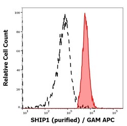
- Separation of MOLT-4 cells (red-filled) from CaCo-2 cells (black-dashed) in flow cytometry analysis (intracellular staining) of cellular suspensions of MOLT-4 and CaCo-2 cell lines stained using anti-SHIP1 (SHIP-01) purified Monoclonal antibody (Product # MA1-10449) (concentration in sample 1 µg/mL, GAM APC).

- Submitted by
- Invitrogen Antibodies (provider)
- Main image

- Experimental details
- Separation of MOLT-4 cells (red-filled) from CaCo-2 cells (black-dashed) in flow cytometry analysis (intracellular staining) of cellular suspensions of MOLT-4 and CaCo-2 cell lines stained using anti-SHIP1 (SHIP-01) purified Monoclonal antibody (Product # MA1-10449) (concentration in sample 1 µg/mL, GAM APC).
