 Explore
 Validate
 Learn
Learn Western blot
Antibody data
- Antibody Data
- Antigen structure
- References [0]
- Comments [0]
- Validations
- Western blot [2]
- Immunocytochemistry [2]
- Flow cytometry [2]
Submit
Validation data
Reference
Comment
Report error
- Product number
- MA1-10450 - Provider product page

- Provider
- Invitrogen Antibodies
- Product name
- SHIP1 Monoclonal Antibody (SHIP-02)
- Antibody type
- Monoclonal
- Antigen
- Synthetic peptide
- Description
- This antibody reacts with SHIP-1, a phosphoinositide phosphatase largely confined to hematopoietic cells. Multiple forms of SHIP-1 have been reported with molecular weights of 110, 125, 130, 135 and 145 kDa. Western Blot: Reducing conditions.
- Reactivity
- Human, Mouse
- Host
- Mouse
- Isotype
- IgG
- Antibody clone number
- SHIP-02
- Vial size
- 100 μg
- Concentration
- 1 mg/mL
- Storage
- 4°C, do not freeze
No comments: Submit comment
Supportive validation
- Submitted by
- Invitrogen Antibodies (provider)
- Main image
- Experimental details
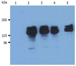
- Western blotting analysis (reducing conditions) of human SHIP-1 in whole cell lysate of THP-1 human acute monocytic leukemia cell line. Lane 1: immunostaining with Isotype mouse IgG1 control (PPV-04). Lane 2,3: immunostaining with anti-human SHIP-1 (SHIP-01) Monoclonal antibody (Product # MA1-10450). Lane 4,5: immunostaining with anti-human SHIP-1 (SHIP-02).

- Submitted by
- Invitrogen Antibodies (provider)
- Main image
- Experimental details
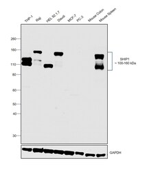
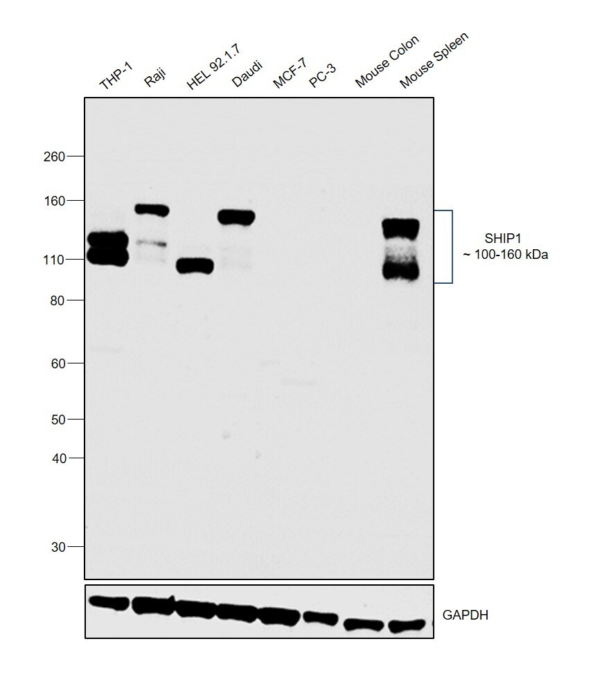
- Western blot was performed using Anti-SHIP1 Monoclonal Antibody (SHIP-02) (Product # MA1-10450) and 100-160 kDa bands corresponding to SHIP1 was observed across cell lines and Mouse Spleen except MCF-7, PC-3 and Mouse Colon which are reported to be low. Membrane enriched extracts (30 µg lysate) of THP-1 (Lane 1), Raji (Lane 2), HEL 92.1.7 (Lane 3), Daudi (Lane 4), MCF-7 (Lane 5), PC-3 (Lane 6), tissue extracts of Mouse Colon (Lane 7) and Mouse Spleen (Lane 8) were electrophoresed using Novex® NuPAGE® 4-12 % Bis-Tris gel (Product # NP0322BOX). Resolved proteins were then transferred onto a nitrocellulose membrane (Product # IB23001) by iBlot® 2 Dry Blotting System (Product # IB21001). The blot was probed with the primary antibody (1:1000 dilution) and detected by chemiluminescence with Goat anti-Mouse IgG (H+L), Superclonal™ Recombinant Secondary Antibody, HRP (Product # A28177, 1:4000 dilution) using the iBright FL 1000 (Product # A32752). Chemiluminescent detection was performed using Novex® ECL Chemiluminescent Substrate Reagent Kit (Product # WP20005).
Supportive validation
- Submitted by
- Invitrogen Antibodies (provider)
- Main image

- Experimental details
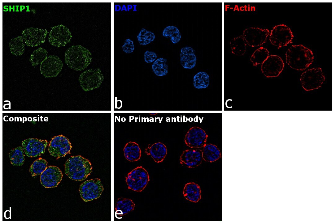
- Immunofluorescence analysis of SHIP1 was performed using 70% confluent log phase THP-1 cells. The cells were fixed with 4% paraformaldehyde for 10 minutes, permeabilized with 0.1% Triton™ X-100 for 15 minutes, and blocked with 2% BSA for 1 hour at room temperature. The cells were labeled with SHIP1 Mouse Monoclonal Antibody (SHIP-02) (Product # MA1-10450) at 1:100 dilution in 0.1% BSA, incubated at 4 degree Celsius overnight and then with Goat anti-Mouse IgG (H+L) Highly Cross-Adsorbed Secondary Antibody, Alexa Fluor Plus 488 (Product # A32723) at a dilution of 1:2000 for 45 minutes at room temperature (Panel a: green). Nuclei (Panel b: blue) were stained with ProLong™ Diamond Antifade Mountant with DAPI (Product # P36962). F-actin (Panel c: red) was stained with Rhodamine Phalloidin (Product # R415, 1:300). Panel d represents the merged image showing membrane and cytoplasmic localization. Panel e represents control cells with no primary antibody to assess background. The images were captured at 60X magnification.
- Submitted by
- Invitrogen Antibodies (provider)
- Main image

- Experimental details
- Immunofluorescence analysis of SHIP1 was performed using 70% confluent log phase THP-1 cells. The cells were fixed with 4% paraformaldehyde for 10 minutes, permeabilized with 0.1% Triton™ X-100 for 15 minutes, and blocked with 2% BSA for 1 hour at room temperature. The cells were labeled with SHIP1 Mouse Monoclonal Antibody (SHIP-02) (Product # MA1-10450) at 1:100 dilution in 0.1% BSA, incubated at 4 degree Celsius overnight and then with Goat anti-Mouse IgG (H+L) Highly Cross-Adsorbed Secondary Antibody, Alexa Fluor Plus 488 (Product # A32723) at a dilution of 1:2000 for 45 minutes at room temperature (Panel a: green). Nuclei (Panel b: blue) were stained with ProLong™ Diamond Antifade Mountant with DAPI (Product # P36962). F-actin (Panel c: red) was stained with Rhodamine Phalloidin (Product # R415, 1:300). Panel d represents the merged image showing membrane and cytoplasmic localization. Panel e represents control cells with no primary antibody to assess background. The images were captured at 60X magnification.

Supportive validation
- Submitted by
- Invitrogen Antibodies (provider)
- Main image

- Experimental details
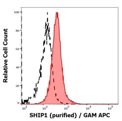
- Separation of MOLT-4 cells stained using anti-SHIP1 (SHIP-02) purified Monoclonal antibody (Product # MA1-10450) (concentration in sample 3 µg/mL, GAM APC, red-filled) from MOLT-4 cells unstained by primary antibody (GAM APC, black-dashed) in flow cytometry analysis (intracellular staining).

- Submitted by
- Invitrogen Antibodies (provider)
- Main image
- Experimental details
- Separation of MOLT-4 cells stained using anti-SHIP1 (SHIP-02) purified Monoclonal antibody (Product # MA1-10450) (concentration in sample 3 µg/mL, GAM APC, red-filled) from MOLT-4 cells unstained by primary antibody (GAM APC, black-dashed) in flow cytometry analysis (intracellular staining).
