Explore
 Validate
 Learn
Learn Western blot
 Immunocytochemistry
Antibody data
- Antibody Data
- Antigen structure
- References [0]
- Comments [0]
- Validations
- Immunocytochemistry [3]
Submit
Validation data
Reference
Comment
Report error
- Product number
- GTX23437 - Provider product page

- Provider
- GeneTex
- Proper citation
- GeneTex Cat#GTX23437, RRID:AB_370005
- Product name
- SAP97 antibody
- Antibody type
- Polyclonal
- Reactivity
- Human, Mouse, Rat
- Host
- Rabbit
No comments: Submit comment
Supportive validation
- Submitted by
- GeneTex (provider)
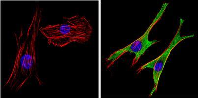
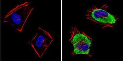
- Main image
- Experimental details
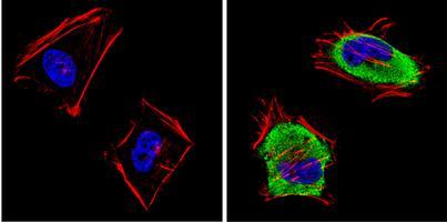
- Immunofluorescent analysis of SAP97 (green) showing staining in the cytoplasm and membrane of C2C12 cells. Formalin-fixed cells were permeabilized with 0.1% Triton X-100 in TBS for 5-10 minutes and blocked with 3% BSA-PBS for 30 minutes at room temperature. Cells were probed with a SAP97 polyclonal antibody (GTX23437) in 3% BSA-PBS at a dilution of 1:100 and incubated overnight at 4 ?C in a humidified chamber. Cells were washed with PBST and incubated with a DyLight-conjugated secondary antibody in PBS at room temperature in the dark. F-actin (red) was stained with a flourescent red phalloidin and nuclei (blue) were stained with Hoechst or DAPI. Images were taken at a magnification of 60x.
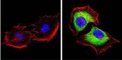
- Submitted by
- GeneTex (provider)
- Main image
- Experimental details
- Immunofluorescent analysis of SAP97 (green) showing staining in the cytoplasm and membrane of C2C12 cells. Formalin-fixed cells were permeabilized with 0.1% Triton X-100 in TBS for 5-10 minutes and blocked with 3% BSA-PBS for 30 minutes at room temperature. Cells were probed with a SAP97 polyclonal antibody (GTX23437) in 3% BSA-PBS at a dilution of 1:100 and incubated overnight at 4 ?C in a humidified chamber. Cells were washed with PBST and incubated with a DyLight-conjugated secondary antibody in PBS at room temperature in the dark. F-actin (red) was stained with a flourescent red phalloidin and nuclei (blue) were stained with Hoechst or DAPI. Images were taken at a magnification of 60x.
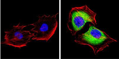
- Submitted by
- GeneTex (provider)
- Main image

- Experimental details
- Immunofluorescent analysis of SAP97 (green) showing staining in the cytoplasm and membrane of C2C12 cells. Formalin-fixed cells were permeabilized with 0.1% Triton X-100 in TBS for 5-10 minutes and blocked with 3% BSA-PBS for 30 minutes at room temperature. Cells were probed with a SAP97 polyclonal antibody (GTX23437) in 3% BSA-PBS at a dilution of 1:100 and incubated overnight at 4 ?C in a humidified chamber. Cells were washed with PBST and incubated with a DyLight-conjugated secondary antibody in PBS at room temperature in the dark. F-actin (red) was stained with a flourescent red phalloidin and nuclei (blue) were stained with Hoechst or DAPI. Images were taken at a magnification of 60x.