Explore
 Validate
 Learn
Learn Western blot
 Immunohistochemistry
Antibody data
- Antibody Data
- Antigen structure
- References [1]
- Comments [0]
- Validations
- Immunohistochemistry [1]
- Flow cytometry [2]
- Other assay [1]
Submit
Validation data
Reference
Comment
Report error
- Product number
- PA5-24581 - Provider product page

- Provider
- Invitrogen Antibodies
- Product name
- TNPO1 Polyclonal Antibody
- Antibody type
- Polyclonal
- Antigen
- Synthetic peptide
- Description
- This antibody is predicted to react with bovine and mouse based on sequence homology.
- Reactivity
- Human
- Host
- Rabbit
- Isotype
- IgG
- Vial size
- 400 μL
- Concentration
- 0.4 mg/mL
- Storage
- Store at 4°C short term. For long term storage, store at -20°C, avoiding freeze/thaw cycles.
Submitted references Active nuclear import of the deacetylase Sirtuin-2 is controlled by its C-terminus and importins.
Eldridge MJG, Pereira JM, Impens F, Hamon MA
Scientific reports 2020 Feb 10;10(1):2034
Scientific reports 2020 Feb 10;10(1):2034
No comments: Submit comment
Supportive validation
- Submitted by
- Invitrogen Antibodies (provider)
- Main image
- Experimental details
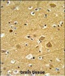
- Immunohistochemistry analysis of TNPO1 in formalin-fixed and paraffin-embedded human brain tissue. Samples were incubated with TNPO1 polyclonal antibody (Product # PA5-24581) which was peroxidase-conjugated to the secondary antibody, followed by DAB staining. This data demonstrates the use of this antibody for immunohistochemistry; clinical relevance has not been evaluated.

Supportive validation
- Submitted by
- Invitrogen Antibodies (provider)
- Main image
- Experimental details
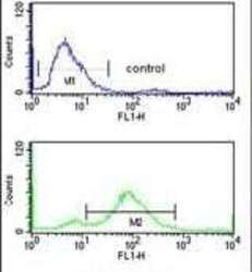
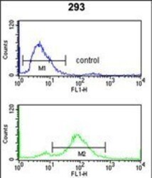
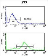
- Flow cytometry analysis of 293 cells using a TNPO1 polyclonal antibody (Product # PA5-24581) (bottom) compared to a negative control cell (top) at a dilution of 1:10-50, followed by a FITC-conjugated goat anti-rabbit antibody
- Submitted by
- Invitrogen Antibodies (provider)
- Main image
- Experimental details
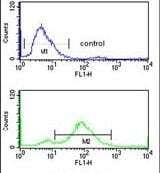
- Flow cytometry of TNPO1 in 293 cells (bottom histogram). Samples were incubated with TNPO1 polyclonal antibody (Product # PA5-24581) followed by FITC-conjugated goat-anti-rabbit secondary antibody. Negative control cell (top histogram).
Supportive validation
- Submitted by
- Invitrogen Antibodies (provider)
- Main image

- Experimental details
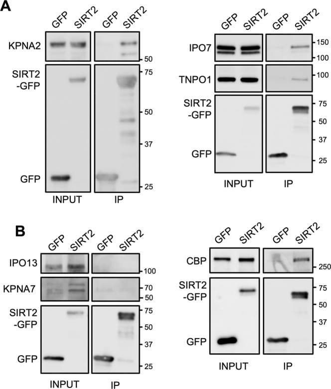
- Figure 2 SIRT2 interacts with multiple importins. SIRT2-GFP or GFP alone where immunoprecipitated using GFP-Trap(r) agarose beads for 1 hr. Cell lysates (INPUT) and IP fractions were immunoblotted using antibodies against GFP and (A) KPNA2, IPO7, TNPO1 or, (B) IPO13 and CBP which served as controls. Images are representative of 3 independent experiments. Uncropped blots are presented in Supplementary S3.