Explore
 Validate
 Learn
Learn Western blot
Antibody data
- Antibody Data
- Antigen structure
- References [4]
- Comments [0]
- Validations
- Western blot [2]
- Immunocytochemistry [2]
- Immunohistochemistry [1]
Submit
Validation data
Reference
Comment
Report error
- Product number
- GTX105038 - Provider product page

- Provider
- GeneTex
- Proper citation
- GeneTex Cat#GTX105038, RRID:AB_2036733
- Product name
- DAG1 antibody
- Antibody type
- Polyclonal
- Reactivity
- Human, Mouse, Horse
- Host
- Rabbit
Submitted references Absence of α- and β-dystroglycan is associated with Walker-Warburg syndrome.
Disease mutations in CMP-sialic acid transporter SLC35A1 result in abnormal α-dystroglycan O-mannosylation, independent from sialic acid.
Transcriptome profiling of granulosa and theca cells during dominant follicle development in the horse.
Virus entry. Lassa virus entry requires a trigger-induced receptor switch.
Riemersma M, Mandel H, van Beusekom E, Gazzoli I, Roscioli T, Eran A, Gershoni-Baruch R, Gershoni M, Pietrokovski S, Vissers LE, Lefeber DJ, Willemsen MA, Wevers RA, van Bokhoven H
Neurology 2015 May 26;84(21):2177-82
Neurology 2015 May 26;84(21):2177-82
Disease mutations in CMP-sialic acid transporter SLC35A1 result in abnormal α-dystroglycan O-mannosylation, independent from sialic acid.
Riemersma M, Sandrock J, Boltje TJ, Büll C, Heise T, Ashikov A, Adema GJ, van Bokhoven H, Lefeber DJ
Human molecular genetics 2015 Apr 15;24(8):2241-6
Human molecular genetics 2015 Apr 15;24(8):2241-6
Transcriptome profiling of granulosa and theca cells during dominant follicle development in the horse.
Donadeu FX, Fahiminiya S, Esteves CL, Nadaf J, Miedzinska K, McNeilly AS, Waddington D, Gérard N
Biology of reproduction 2014 Nov;91(5):111
Biology of reproduction 2014 Nov;91(5):111
Virus entry. Lassa virus entry requires a trigger-induced receptor switch.
Jae LT, Raaben M, Herbert AS, Kuehne AI, Wirchnianski AS, Soh TK, Stubbs SH, Janssen H, Damme M, Saftig P, Whelan SP, Dye JM, Brummelkamp TR
Science (New York, N.Y.) 2014 Jun 27;344(6191):1506-10
Science (New York, N.Y.) 2014 Jun 27;344(6191):1506-10
No comments: Submit comment
Supportive validation
- Submitted by
- GeneTex (provider)
- Main image
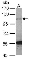
- Experimental details
- Sample (30 ug of whole cell lysate) A: U87-MG 7.5% SDS PAGE GTX105038 diluted at 1:500
- Validation comment
- WB

- Submitted by
- GeneTex (provider)
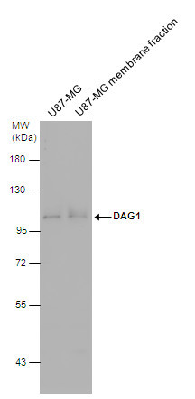
- Main image
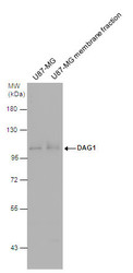
- Experimental details
- DAG1 antibody detects DAG1 protein by western blot analysis. U87-MG whole cell extracts and membrane extracts (30 ?g) were separated by 7.5% SDS-PAGE, and the membrane was blotted with DAG1 antibody (GTX105038) diluted at 1:500. The HRP-conjugated anti-rabbit IgG antibody (GTX213110-01) was used to detect the primary antibody.
Supportive validation
- Submitted by
- GeneTex (provider)
- Main image
- Experimental details
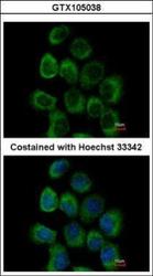
- Immunofluorescence analysis of paraformaldehyde-fixed A431, using alpha Dystroglycan(GTX105038) antibody at 1:500 dilution.

- Submitted by
- GeneTex (provider)
- Main image
- Experimental details
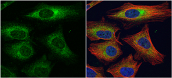
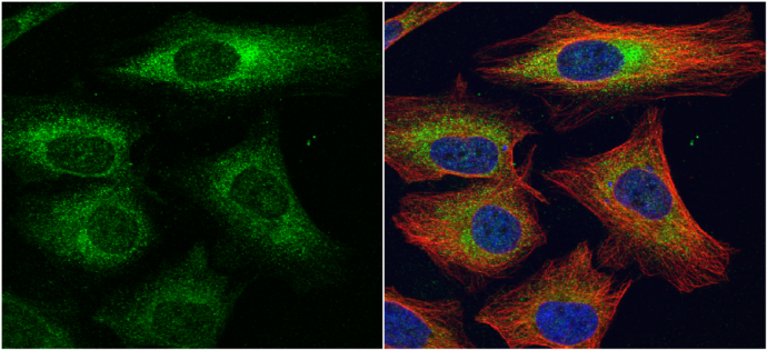
- alpha Dystroglycan antibody detects alpha Dystroglycan protein at cytoplasm by immunofluorescent analysis.Sample: HeLa cells were fixed in 4% paraformaldehyde at RT for 15 min.Green: alpha Dystroglycan protein stained by alpha Dystroglycan antibody (GTX105038) diluted at 1:1000.Red: alpha Tubulin, a cytoskeleton marker, stained by alpha Tubulin antibody [B-5-1-2] (GTX11304) diluted at 1:10000.Blue: Hoechst 33342 staining.
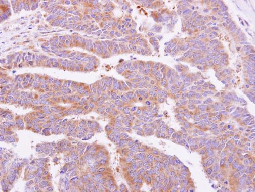
Supportive validation
- Submitted by
- GeneTex (provider)
- Main image

- Experimental details
- alpha Dystroglycan antibody detects DAG1 protein at cytoplasm on human colon carcinoma by immunohistochemical analysis. Sample: Paraffin-embedded colon carcinoma. alpha Dystroglycan antibody (GTX105038) dilution: 1:500.