 Explore
 Validate
 Learn
Learn Western blot
Antibody data
- Antibody Data
- Antigen structure
- References [0]
- Comments [0]
- Validations
- Western blot [5]
- Immunocytochemistry [1]
- Immunohistochemistry [1]
Submit
Validation data
Reference
Comment
Report error
- Product number
- PA5-34908 - Provider product page

- Provider
- Invitrogen Antibodies
- Product name
- beta Dystroglycan Polyclonal Antibody
- Antibody type
- Polyclonal
- Antigen
- Recombinant protein fragment
- Description
- Recommended positive controls: H1299, MCF-7. Predicted reactivity: Mouse (89%), Rat (87%), Dog (89%), Cat (94%), Rabbit (89%), Rhesus Monkey (97%), Bovine (91%). Store product as a concentrated solution. Centrifuge briefly prior to opening the vial.
- Reactivity
- Human, Mouse
- Host
- Rabbit
- Isotype
- IgG
- Vial size
- 100 µL
- Concentration
- 0.6 mg/mL
- Storage
- Store at 4°C short term. For long term storage, store at -20°C, avoiding freeze/thaw cycles.
No comments: Submit comment
Supportive validation
- Submitted by
- Invitrogen Antibodies (provider)
- Main image
- Experimental details
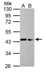
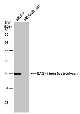
- Western blot analysis of beta Dystroglycan using 30 µg of A) H1299 and B) MCF-7 lysate. Samples were loaded onto a 10% SDS-PAGE gel and probed with a beta Dystroglycan polyclonal antibody (Product # PA5-34908) at a dilution of 1:2000.

- Submitted by
- Invitrogen Antibodies (provider)
- Main image
- Experimental details
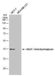
- Western Blot analysis of beta Dystroglycan was performed by separating 30 µg of various whole cell extracts by 10% SDS-PAGE. Proteins were transferred to a membrane and probed with a beta Dystroglycan Polyclonal Antibody (Product # PA5-34908) at a dilution of 1:2000 and a HRP-conjugated anti-rabbit IgG secondary antibody.
- Submitted by
- Invitrogen Antibodies (provider)
- Main image
- Experimental details
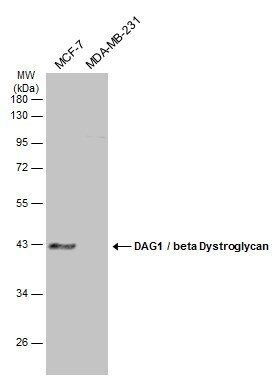
- Western Blot using beta Dystroglycan Polyclonal Antibody (Product # PA5-34908). Various whole cell extracts (30 µg) were separated by 10% SDS-PAGE, and the membrane was blotted with DAG1/beta Dystroglycan antibody [C2C3-2], C-term beta Dystroglycan Polyclonal Antibody (Product # PA5-34908) diluted at 1:2,000. The HRP-conjugated anti-rabbit IgG antibody was used to detect the primary antibody.
- Submitted by
- Invitrogen Antibodies (provider)
- Main image
- Experimental details
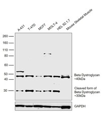
- Western blot was performed using Anti-beta Dystroglycan Polyclonal Antibody (Product # PA5-34908) and a 40kDa band corresponding to beta dystroglycan was observed across all cell lines and tissues tested. A 30kDa cleaved form of beta dystroglycan was also observed in all cell lines. Whole cell extracts (30 µg lysate) of A-431 (Lane 1), T-47D (Lane 2), MCF7 (Lane 3), MOLT-4 (Lane 4), HEL 92.1.7 (Lane 5) and Mouse Skeletal Muscle (Lane 6) were electrophoresed using Novex® NuPAGE® 4-12 % Bis-Tris gel (Product # NP0322BOX). Resolved proteins were then transferred onto a nitrocellulose membrane (Product # IB23001) by iBlot® 2 Dry Blotting System (Product # IB21001). The blot was probed with the primary antibody (1:1000 dilution) and detected by chemiluminescence with Goat anti-Rabbit IgG (H+L), Superclonal™ Recombinant Secondary Antibody, HRP (Product # A27036, 1:4000 dilution) using the iBright FL 1000 (Product # A32752). Chemiluminescent detection was performed using Novex® ECL Chemiluminescent Substrate Reagent Kit (Product # WP20005).

- Submitted by
- Invitrogen Antibodies (provider)
- Main image
- Experimental details
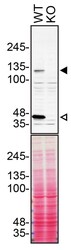
- Western blot of Dystroglycan 1 was performed by loading 90 µg of WT (lane 1) and DAG1 CRISPR KO (lane 2) A431 cell lysates in RIPA buffer onto a 3-12% gradient polyacrylamide gel. Proteins on the blots were visualized with Ponceau staining (below immunoblot). Proteins were transferred to nitrocellulose membrane and blocked in 5% milk for 1 hr. Full-length DAG1 (designated by the black arrow) and beta-dystroglycan (designated by the white arrow) was detected using a DAG1 polyclonal antibody (Product # PA5-34908) at a dilution of 1:500 in 5% BSA in TBS with 0.1% Tween 20 (TBST) overnight at 4°C. The peroxidase-conjugated secondary antibody (Product # 65-6120) was diluted to 0.2 µg/mL in TBST with 5% milk for 1 hr. Chemiluminescent detection was performed using Pierce ECL Western Blotting Substrate (Product # 32106). Data courtesy of YCharOS Inc., an open science company with the mission of characterizing commercially available antibodies using knockout validation.

Supportive validation
- Submitted by
- Invitrogen Antibodies (provider)
- Main image
- Experimental details
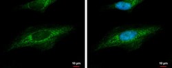
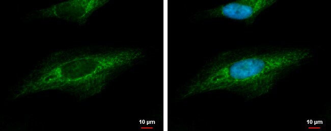
- beta dystroglycan antibody [C2C3-2], C-term detects beta dystroglycan protein at cytoskeleton by immunofluorescent analysis. Sample: HeLa cells were fixed in 4% paraformaldehyde at RT for 15 min. Green: beta dystroglycan protein stained by beta dystroglycan antibody [C2C3-2], C-term (Product # PA5-34908) diluted at 1:500. Blue: Hoechst 33342 staining.
Supportive validation
- Submitted by
- Invitrogen Antibodies (provider)
- Main image
- Experimental details
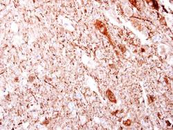
- beta dystroglycan antibody [C2C3-2], C-term detects beta dystroglycan protein at cytosol on mouse brain stem by immunohistochemical analysis. Sample: Paraffin-embedded mouse brain stem. Beta dystroglycan antibody [C2C3-2], C-term (Product # PA5-34908) dilution: 1:250. Antigen Retrieval: EDTA based buffer, pH 8.0, 15 min.
