Explore
 Validate
 Learn
Learn Western blot
Antibody data
- Antibody Data
- Antigen structure
- References [2]
- Comments [0]
- Validations
- Western blot [2]
- Immunohistochemistry [1]
Submit
Validation data
Reference
Comment
Report error
- Product number
- AF6868 - Provider product page

- Provider
- R&D Systems
- Product name
- Human Dystroglycan Antibody
- Antibody type
- Polyclonal
- Description
- Antigen Affinity-purified. Detects human Dystroglycan in direct ELISAs and Western blots.
- Reactivity
- Human
- Host
- Sheep
- Conjugate
- Unconjugated
- Antigen sequence
Q14118- Isotype
- IgG
- Vial size
- 100 ug
- Concentration
- LYOPH
- Storage
- Use a manual defrost freezer and avoid repeated freeze-thaw cycles. 12 months from date of receipt, -20 to -70 °C as supplied. 1 month, 2 to 8 °C under sterile conditions after reconstitution. 6 months, -20 to -70 °C under sterile conditions after reconstitution.
Submitted references Ribitol restores functionally glycosylated α-dystroglycan and improves muscle function in dystrophic FKRP-mutant mice.
Structural basis of laminin binding to the LARGE glycans on dystroglycan.
Cataldi MP, Lu P, Blaeser A, Lu QL
Nature communications 2018 Aug 27;9(1):3448
Nature communications 2018 Aug 27;9(1):3448
Structural basis of laminin binding to the LARGE glycans on dystroglycan.
Briggs DC, Yoshida-Moriguchi T, Zheng T, Venzke D, Anderson ME, Strazzulli A, Moracci M, Yu L, Hohenester E, Campbell KP
Nature chemical biology 2016 Oct;12(10):810-4
Nature chemical biology 2016 Oct;12(10):810-4
No comments: Submit comment
Supportive validation
- Submitted by
- R&D Systems (provider)
- Main image

- Experimental details
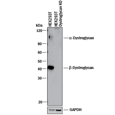
- Western Blot Shows Human Dystroglycan Specificity by Using Knockout Cell Line. Western blot shows lysates of HEK293T human embryonic kidney parental cell line and Dystroglycan knockout HEK293T cell line (KO). PVDF membrane was probed with 1 µg/mL of Sheep Anti-Human Dystroglycan Antigen Affinity-purified Polyclonal Antibody (Catalog # AF6868) followed by HRP-conjugated Anti-Sheep IgG Secondary Antibody (Catalog # HAF016). Specific bands were detected for alpha--Dystroglycan at approximately 110 kDa -and beta-Dystroglycan at approximately 42 kDa (as indicated) in the parental HEK293T cell line, but is not detectable in knockout HEK293T cell line. GAPDH (Catalog # AF5718) is shown as a loading control. This experiment was conducted under reducing conditions and using Immunoblot Buffer Group 1.
- Submitted by
- R&D Systems (provider)
- Main image

- Experimental details
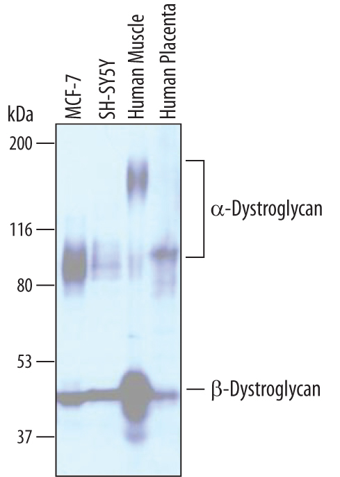
- Detection of Human Dystroglycan by Western Blot. Western blot shows lysates of MCF-7 human breast cancer cell line, SH-SY5Y human neuroblastoma cell line, human muscle tissue, and human placenta tissue. PVDF membrane was probed with 1 µg/mL of Sheep Anti-Human Dystroglycan Antigen Affinity-purified Polyclonal Antibody (Catalog # AF6868) followed by HRP-conjugated Anti-Sheep IgG Secondary Antibody (Catalog # HAF016). Specific bands were detected for alpha-Dystroglycan at approximately 100-160 kDa (as indicated) and beta-Dystroglycan at approximately 42-44 kDa (as indicated). This experiment was conducted under reducing conditions and using Immunoblot Buffer Group 1.
Supportive validation
- Submitted by
- R&D Systems (provider)
- Main image

- Experimental details
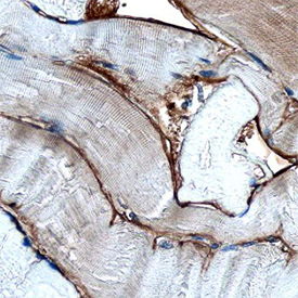
- Dystroglycan in Human Skeletal Muscle. Dystroglycan was detected in immersion fixed paraffin-embedded sections of human skeletal muscle using Sheep Anti-Human Dystroglycan Antigen Affinity-purified Polyclonal Antibody (Catalog # AF6868) at 3 µg/mL overnight at 4 °C. Before incubation with the primary antibody, tissue was subjected to heat-induced epitope retrieval using Antigen Retrieval Reagent-Basic (Catalog # CTS013). Tissue was stained using the Anti-Sheep HRP-DAB Cell & Tissue Staining Kit (brown; Catalog # CTS019) and counterstained with hematoxylin (blue). Specific staining was localized to basement membrane. View our protocol for Chromogenic IHC Staining of Paraffin-embedded Tissue Sections.