 Explore
 Validate
 Learn
Learn Western blot
 Other assay
Antibody data
- Antibody Data
- Antigen structure
- References [35]
- Comments [0]
- Validations
- Other assay [9]
Submit
Validation data
Reference
Comment
Report error
- Product number
- AHP0022 - Provider product page

- Provider
- Invitrogen Antibodies
- Product name
- Aggrecan Monoclonal Antibody (969D4D11)
- Antibody type
- Monoclonal
- Antigen
- Purifed from natural sources
- Description
- This product is recommended as a detection antibody in Sandwich ELISA applications. This clone can also be referred to as clone number 969D 4D11 2A9.
- Reactivity
- Human
- Host
- Mouse
- Isotype
- IgG
- Antibody clone number
- 969D4D11
- Vial size
- 500 µL
- Concentration
- 1 mg/mL
- Storage
- 4° C
Submitted references Associations Between Physical Activity, Self-reported Joint Function, and Molecular Biomarkers in Working Age Individuals With Hip and/or Knee Osteoarthritis.
Characterization of Migratory Cells From Bioengineered Bovine Cartilage in a 3D Co-culture Model.
Utility of direct 3D co-culture model for chondrogenic differentiation of mesenchymal stem cells on hyaluronan scaffold (Hyaff-11).
Platelet-rich plasma enhances the integration of bioengineered cartilage with native tissue in an in vitro model.
Surgical reconstruction of ruptured anterior cruciate ligament prolongs trauma-induced increase of inflammatory cytokines in synovial fluid: an exploratory analysis in the KANON trial.
ESTABLISHING A LIVE CARTILAGE-ON-CARTILAGE INTERFACE FOR TRIBOLOGICAL TESTING.
Formation of Hyaline Cartilage Tissue by Passaged Human Osteoarthritic Chondrocytes.
Generating Mechanically Stable, Pediatric, and Scaffold-Free Nasal Cartilage Constructs In Vitro.
Inflammatory cytokines and biomarkers of cartilage metabolism 8 years after anterior cruciate ligament reconstruction: results from operated and contralateral knees.
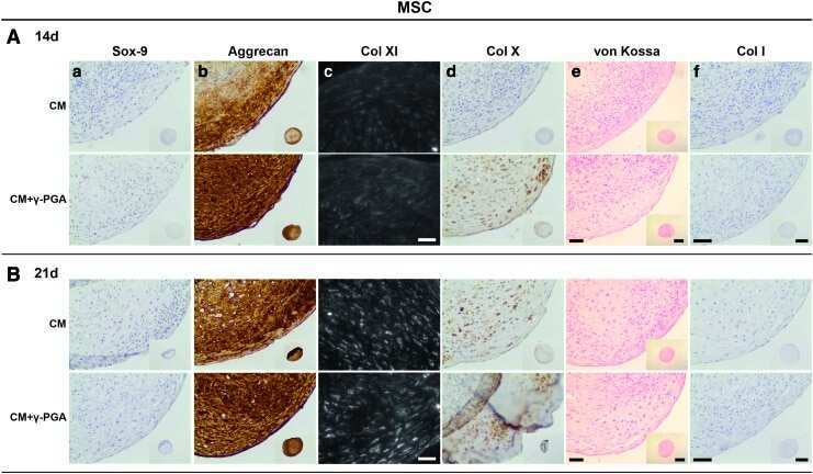
Poly(γ-Glutamic Acid) as an Exogenous Promoter of Chondrogenic Differentiation of Human Mesenchymal Stem/Stromal Cells.
The importance of connexin hemichannels during chondroprogenitor cell differentiation in hydrogel versus microtissue culture models.
Translational development of an ADAMTS-5 antibody for osteoarthritis disease modification.
Biochemical and structural characterization of neocartilage formed by mesenchymal stem cells in alginate hydrogels.
Hyaluronic acid enhances the mechanical properties of tissue-engineered cartilage constructs.
Physiological testosterone levels enhance chondrogenic extracellular matrix synthesis by male intervertebral disc cells in vitro, but not by mesenchymal stem cells.
An ARGS-aggrecan assay for analysis in blood and synovial fluid.
Degenerated human intervertebral discs contain autoantibodies against extracellular matrix proteins.
Similar properties of chondrocytes from osteoarthritis joints and mesenchymal stem cells from healthy donors for tissue engineering of articular cartilage.
Not all lubricin isoforms are substituted with a glycosaminoglycan chain.
Cartilage and bone markers and inflammatory cytokines are increased in synovial fluid in the acute phase of knee injury (hemarthrosis)--a cross-sectional analysis.
The association between changes in synovial fluid levels of ARGS-aggrecan fragments, progression of radiographic osteoarthritis and self-reported outcomes: a cohort study.
The cartilage matrix molecule components produced by human foetal cartilage rudiment cells within scaffolds and the role of exogenous growth factors.
Epiphyseal Chondroprogenitors Provide a Stable Cell Source for Cartilage Cell Therapy.
Influence of different commercial scaffolds on the in vitro differentiation of human mesenchymal stem cells to nucleus pulposus-like cells.
Association between synovial fluid levels of aggrecan ARGS fragments and radiographic progression in knee osteoarthritis.
Changes in serum and synovial fluid biomarkers after acute injury (NCT00332254).
A short-term pharmacodynamic model for monitoring aggrecanase activity: injection of monosodium iodoacetate (MIA) in rats and assessment of aggrecan neoepitope release in synovial fluid using novel ELISAs.
Development of a novel clinical biomarker assay to detect and quantify aggrecanase-generated aggrecan fragments in human synovial fluid, serum and urine.
Comparative immunolocalisation of perlecan with collagen II and aggrecan in human foetal, newborn and adult ovine joint tissues demonstrates perlecan as an early developmental chondrogenic marker.
Silkworm and spider silk scaffolds for chondrocyte support.
The use of Histochoice for histological examination of articular and growth plate cartilages, intervertebral disc and meniscus.
The structure, location, and function of perlecan, a prominent pericellular proteoglycan of fetal, postnatal, and mature hyaline cartilages.
Perlecan displays variable spatial and temporal immunolocalisation patterns in the articular and growth plate cartilages of the ovine stifle joint.
Characterisation of human knee meniscus cell phenotype.
Culture of chondrocytes in alginate surrounded by fibrin gel: characteristics of the cells over a period of eight weeks.
Östlind E, Eek F, Stigmar K, Sant'Anna A, Ekvall Hansson E, Struglics A
Clinical medicine insights. Arthritis and musculoskeletal disorders 2022;15:11795441221081063
Clinical medicine insights. Arthritis and musculoskeletal disorders 2022;15:11795441221081063
Characterization of Migratory Cells From Bioengineered Bovine Cartilage in a 3D Co-culture Model.
Wu MJM, Sermer C, Kandel RA, Theodoropoulos JS
The American journal of sports medicine 2022 Sep;50(11):3090-3101
The American journal of sports medicine 2022 Sep;50(11):3090-3101
Utility of direct 3D co-culture model for chondrogenic differentiation of mesenchymal stem cells on hyaluronan scaffold (Hyaff-11).
Deszcz I, Lis-Nawara A, Grelewski P, Dragan S, Bar J
Regenerative biomaterials 2020 Dec;7(6):543-552
Regenerative biomaterials 2020 Dec;7(6):543-552
Platelet-rich plasma enhances the integration of bioengineered cartilage with native tissue in an in vitro model.
Sermer C, Kandel R, Anderson J, Hurtig M, Theodoropoulos J
Journal of tissue engineering and regenerative medicine 2018 Feb;12(2):427-436
Journal of tissue engineering and regenerative medicine 2018 Feb;12(2):427-436
Surgical reconstruction of ruptured anterior cruciate ligament prolongs trauma-induced increase of inflammatory cytokines in synovial fluid: an exploratory analysis in the KANON trial.
Larsson S, Struglics A, Lohmander LS, Frobell R
Osteoarthritis and cartilage 2017 Sep;25(9):1443-1451
Osteoarthritis and cartilage 2017 Sep;25(9):1443-1451
ESTABLISHING A LIVE CARTILAGE-ON-CARTILAGE INTERFACE FOR TRIBOLOGICAL TESTING.
Trevino RL, Stoia J, Laurent MP, Pacione CA, Chubinskaya S, Wimmer MA
Biotribology (Oxford) 2017 Mar;9:1-11
Biotribology (Oxford) 2017 Mar;9:1-11
Formation of Hyaline Cartilage Tissue by Passaged Human Osteoarthritic Chondrocytes.
Bianchi VJ, Weber JF, Waldman SD, Backstein D, Kandel RA
Tissue engineering. Part A 2017 Feb;23(3-4):156-165
Tissue engineering. Part A 2017 Feb;23(3-4):156-165
Generating Mechanically Stable, Pediatric, and Scaffold-Free Nasal Cartilage Constructs In Vitro.
Akbari P, Waldman SD, Propst EJ, Cushing SL, Weber JF, Yeger H, Farhat WA
Tissue engineering. Part C, Methods 2016 Dec;22(12):1077-1084
Tissue engineering. Part C, Methods 2016 Dec;22(12):1077-1084
Inflammatory cytokines and biomarkers of cartilage metabolism 8 years after anterior cruciate ligament reconstruction: results from operated and contralateral knees.
Åhlén M, Roshani L, Lidén M, Struglics A, Rostgård-Christensen L, Kartus J
The American journal of sports medicine 2015 Jun;43(6):1460-6
The American journal of sports medicine 2015 Jun;43(6):1460-6
Poly(γ-Glutamic Acid) as an Exogenous Promoter of Chondrogenic Differentiation of Human Mesenchymal Stem/Stromal Cells.
Antunes JC, Tsaryk R, Gonçalves RM, Pereira CL, Landes C, Brochhausen C, Ghanaati S, Barbosa MA, Kirkpatrick CJ
Tissue engineering. Part A 2015 Jun;21(11-12):1869-85
Tissue engineering. Part A 2015 Jun;21(11-12):1869-85
The importance of connexin hemichannels during chondroprogenitor cell differentiation in hydrogel versus microtissue culture models.
Schrobback K, Klein TJ, Woodfield TB
Tissue engineering. Part A 2015 Jun;21(11-12):1785-94
Tissue engineering. Part A 2015 Jun;21(11-12):1785-94
Translational development of an ADAMTS-5 antibody for osteoarthritis disease modification.
Larkin J, Lohr TA, Elefante L, Shearin J, Matico R, Su JL, Xue Y, Liu F, Genell C, Miller RE, Tran PB, Malfait AM, Maier CC, Matheny CJ
Osteoarthritis and cartilage 2015 Aug;23(8):1254-66
Osteoarthritis and cartilage 2015 Aug;23(8):1254-66
Biochemical and structural characterization of neocartilage formed by mesenchymal stem cells in alginate hydrogels.
Olderøy MØ, Lilledahl MB, Beckwith MS, Melvik JE, Reinholt F, Sikorski P, Brinchmann JE
PloS one 2014;9(3):e91662
PloS one 2014;9(3):e91662
Hyaluronic acid enhances the mechanical properties of tissue-engineered cartilage constructs.
Levett PA, Hutmacher DW, Malda J, Klein TJ
PloS one 2014;9(12):e113216
PloS one 2014;9(12):e113216
Physiological testosterone levels enhance chondrogenic extracellular matrix synthesis by male intervertebral disc cells in vitro, but not by mesenchymal stem cells.
Bertolo A, Baur M, Aebli N, Ferguson SJ, Stoyanov J
The spine journal : official journal of the North American Spine Society 2014 Mar 1;14(3):455-68
The spine journal : official journal of the North American Spine Society 2014 Mar 1;14(3):455-68
An ARGS-aggrecan assay for analysis in blood and synovial fluid.
Larsson S, Lohmander LS, Struglics A
Osteoarthritis and cartilage 2014 Feb;22(2):242-9
Osteoarthritis and cartilage 2014 Feb;22(2):242-9
Degenerated human intervertebral discs contain autoantibodies against extracellular matrix proteins.
Capossela S, Schläfli P, Bertolo A, Janner T, Stadler BM, Pötzel T, Baur M, Stoyanov JV
European cells & materials 2014 Apr 4;27:251-63; discussion 263
European cells & materials 2014 Apr 4;27:251-63; discussion 263
Similar properties of chondrocytes from osteoarthritis joints and mesenchymal stem cells from healthy donors for tissue engineering of articular cartilage.
Fernandes AM, Herlofsen SR, Karlsen TA, Küchler AM, Fløisand Y, Brinchmann JE
PloS one 2013;8(5):e62994
PloS one 2013;8(5):e62994
Not all lubricin isoforms are substituted with a glycosaminoglycan chain.
Lord MS, Estrella RP, Chuang CY, Youssef P, Karlsson NG, Flannery CR, Whitelock JM
Connective tissue research 2012;53(2):132-41
Connective tissue research 2012;53(2):132-41
Cartilage and bone markers and inflammatory cytokines are increased in synovial fluid in the acute phase of knee injury (hemarthrosis)--a cross-sectional analysis.
Swärd P, Frobell R, Englund M, Roos H, Struglics A
Osteoarthritis and cartilage 2012 Nov;20(11):1302-8
Osteoarthritis and cartilage 2012 Nov;20(11):1302-8
The association between changes in synovial fluid levels of ARGS-aggrecan fragments, progression of radiographic osteoarthritis and self-reported outcomes: a cohort study.
Larsson S, Englund M, Struglics A, Lohmander LS
Osteoarthritis and cartilage 2012 May;20(5):388-395
Osteoarthritis and cartilage 2012 May;20(5):388-395
The cartilage matrix molecule components produced by human foetal cartilage rudiment cells within scaffolds and the role of exogenous growth factors.
Chuang CY, Shahin K, Lord MS, Melrose J, Doran PM, Whitelock JM
Biomaterials 2012 Jun;33(16):4078-88
Biomaterials 2012 Jun;33(16):4078-88
Epiphyseal Chondroprogenitors Provide a Stable Cell Source for Cartilage Cell Therapy.
Darwiche S, Scaletta C, Raffoul W, Pioletti DP, Applegate LA
Cell medicine 2012 Jan;4(1):23-32
Cell medicine 2012 Jan;4(1):23-32
Influence of different commercial scaffolds on the in vitro differentiation of human mesenchymal stem cells to nucleus pulposus-like cells.
Bertolo A, Mehr M, Aebli N, Baur M, Ferguson SJ, Stoyanov JV
European spine journal : official publication of the European Spine Society, the European Spinal Deformity Society, and the European Section of the Cervical Spine Research Society 2012 Aug;21 Suppl 6(Suppl 6):S826-38
European spine journal : official publication of the European Spine Society, the European Spinal Deformity Society, and the European Section of the Cervical Spine Research Society 2012 Aug;21 Suppl 6(Suppl 6):S826-38
Association between synovial fluid levels of aggrecan ARGS fragments and radiographic progression in knee osteoarthritis.
Larsson S, Englund M, Struglics A, Lohmander LS
Arthritis research & therapy 2010;12(6):R230
Arthritis research & therapy 2010;12(6):R230
Changes in serum and synovial fluid biomarkers after acute injury (NCT00332254).
Catterall JB, Stabler TV, Flannery CR, Kraus VB
Arthritis research & therapy 2010;12(6):R229
Arthritis research & therapy 2010;12(6):R229
A short-term pharmacodynamic model for monitoring aggrecanase activity: injection of monosodium iodoacetate (MIA) in rats and assessment of aggrecan neoepitope release in synovial fluid using novel ELISAs.
Swearingen CA, Chambers MG, Lin C, Marimuthu J, Rito CJ, Carter QL, Dotzlaf J, Liu C, Chandrasekhar S, Duffin KL, Mitchell PG, Durham TB, Wiley MR, Thirunavukkarasu K
Osteoarthritis and cartilage 2010 Sep;18(9):1159-66
Osteoarthritis and cartilage 2010 Sep;18(9):1159-66
Development of a novel clinical biomarker assay to detect and quantify aggrecanase-generated aggrecan fragments in human synovial fluid, serum and urine.
Swearingen CA, Carpenter JW, Siegel R, Brittain IJ, Dotzlaf J, Durham TB, Toth JL, Laska DA, Marimuthu J, Liu C, Brown DP, Carter QL, Wiley MR, Duffin KL, Mitchell PG, Thirunavukkarasu K
Osteoarthritis and cartilage 2010 Sep;18(9):1150-8
Osteoarthritis and cartilage 2010 Sep;18(9):1150-8
Comparative immunolocalisation of perlecan with collagen II and aggrecan in human foetal, newborn and adult ovine joint tissues demonstrates perlecan as an early developmental chondrogenic marker.
Smith SM, Shu C, Melrose J
Histochemistry and cell biology 2010 Sep;134(3):251-63
Histochemistry and cell biology 2010 Sep;134(3):251-63
Silkworm and spider silk scaffolds for chondrocyte support.
Gellynck K, Verdonk PC, Van Nimmen E, Almqvist KF, Gheysens T, Schoukens G, Van Langenhove L, Kiekens P, Mertens J, Verbruggen G
Journal of materials science. Materials in medicine 2008 Nov;19(11):3399-409
Journal of materials science. Materials in medicine 2008 Nov;19(11):3399-409
The use of Histochoice for histological examination of articular and growth plate cartilages, intervertebral disc and meniscus.
Melrose J, Smith SM, Smith MM, Little CB
Biotechnic & histochemistry : official publication of the Biological Stain Commission 2008 Feb;83(1):47-53
Biotechnic & histochemistry : official publication of the Biological Stain Commission 2008 Feb;83(1):47-53
The structure, location, and function of perlecan, a prominent pericellular proteoglycan of fetal, postnatal, and mature hyaline cartilages.
Melrose J, Roughley P, Knox S, Smith S, Lord M, Whitelock J
The Journal of biological chemistry 2006 Dec 1;281(48):36905-14
The Journal of biological chemistry 2006 Dec 1;281(48):36905-14
Perlecan displays variable spatial and temporal immunolocalisation patterns in the articular and growth plate cartilages of the ovine stifle joint.
Melrose J, Smith S, Cake M, Read R, Whitelock J
Histochemistry and cell biology 2005 Jun;123(6):561-71
Histochemistry and cell biology 2005 Jun;123(6):561-71
Characterisation of human knee meniscus cell phenotype.
Verdonk PC, Forsyth RG, Wang J, Almqvist KF, Verdonk R, Veys EM, Verbruggen G
Osteoarthritis and cartilage 2005 Jul;13(7):548-60
Osteoarthritis and cartilage 2005 Jul;13(7):548-60
Culture of chondrocytes in alginate surrounded by fibrin gel: characteristics of the cells over a period of eight weeks.
Almqvist KF, Wang L, Wang J, Baeten D, Cornelissen M, Verdonk R, Veys EM, Verbruggen G
Annals of the rheumatic diseases 2001 Aug;60(8):781-90
Annals of the rheumatic diseases 2001 Aug;60(8):781-90
No comments: Submit comment
Supportive validation
- Submitted by
- Invitrogen Antibodies (provider)
- Main image
- Experimental details
- NULL
- Submitted by
- Invitrogen Antibodies (provider)
- Main image
- Experimental details
- NULL

- Submitted by
- Invitrogen Antibodies (provider)
- Main image
- Experimental details
- NULL
- Submitted by
- Invitrogen Antibodies (provider)
- Main image
- Experimental details
- NULL

- Submitted by
- Invitrogen Antibodies (provider)
- Main image

- Experimental details
- NULL
- Submitted by
- Invitrogen Antibodies (provider)
- Main image
- Experimental details
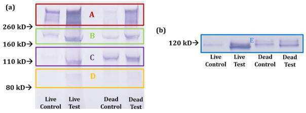
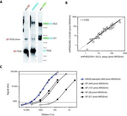
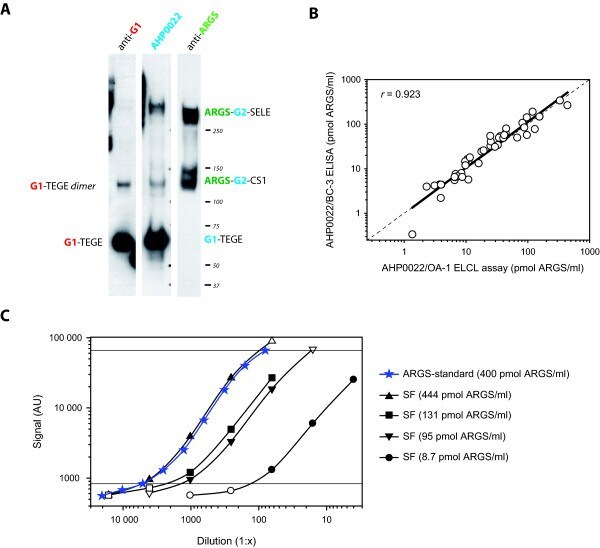
- Figure 2 Specificity and technical performance of the aggrecan capture OA-1 ARGS ELCL assay . (a) 2.4 mug of ADAMTS-4-digested and deglycosylated human aggrecan was separated on a 3 to 8% Tris-acetate gel, transferred and probed with anti-G1, anti-aggrecan (AHP0022), or anti-ARGS (OA-1). Protein standard molecular weights and aggrecan fragments detected are indicated. Epitopes recognized by the antibodies are written in color: red, anti-G1; blue, anti-aggrecan (AHP0022); green, anti-ARGS. Aggrecan domains and amino acid sequences: G1 and G2, globular domains 1 and 2; CS1 and CS2, chondroitin sulfate-rich domains 1 and 2; TEGE and ARGS: C- and N-terminal amino acid sequences at the aggrecanase cleavage site within the interglobular domain; SELE, C-terminal amino acid sequence of aggrecanase cleavage within the CS2 [ 1 ]. (b) ARGS concentration measured with the OA-1 ARGS ELCL assay and the BC-3 ARGS ELISA [ 35 ] in 43 individual SFs with a regression line, dashed line of equality, and Pearson's correlation coefficient ( r ). (c) Dilution curves of ARGS-aggrecan standard and four synovial fluids (SFs) analyzed in the OA-1 ARGS ELCL assay. Sample concentrations of ARGS-aggrecan at different dilutions were calculated from the standard curve (four-parameter logistic). Dilutions falling within the range of detection of the standard curve (solid symbols within horizontal lines) were used for linearity of dilution calculations (Table 2), with mean values presented in the legend.
- Submitted by
- Invitrogen Antibodies (provider)
- Main image
- Experimental details
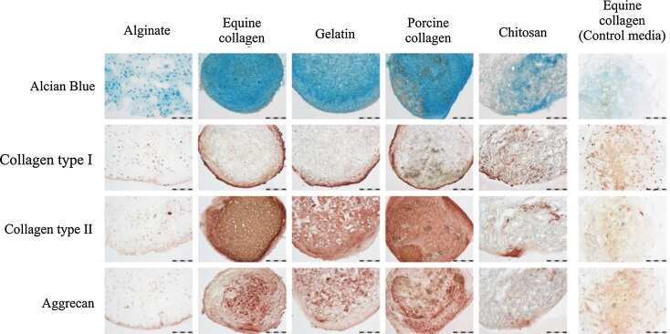
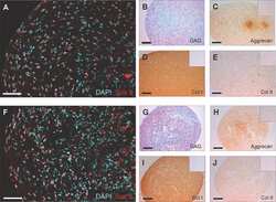
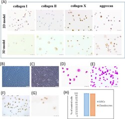
- Figure 1 Immunophenotypic and morphological features of MSCs and chondrocytes in direct co-culture. (A) Immunohistochemical staining of type I, II, X collagen and aggrecan in MSCs/chondrocytes after 17 days of differentiation in direct 2D and 3D co-culture model (EnVision technique, scale bar = 50 um); (B) Chondrocytes and (G) direct co-culture on culture flask (inverted phase contrast microscope, scale bar = 100 um); (C) Hematoxylin-eosin staining of chondroinduced MSCs and (D) co-culture in 2D culture after 14 days (scale bar = 50 um); (E) Expression of vimentin in MSCs/chondrocytes and (F) chondroinduced MSCs after 17 days of culture in direct 2D co-culture model (EnVision technique, scale bar = 50 um); (H) The viability of MSCs and chondrocytes on the Hyaff-11 membrane after 48 h. Data are given as mean +- standard deviation.
- Submitted by
- Invitrogen Antibodies (provider)
- Main image
- Experimental details
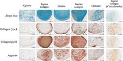
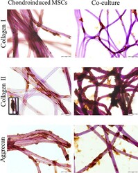
- Figure 2 Immunohistochemical staining of type I, II collagen and aggrecan in MSCs in standard culture and MSCs/chondrocytes in direct co-culture model after 17 days differentiation on Hyaff-11 membrane (EnVision technique, scale bar = 50 um, co-culture collagen I scale bar = 100 um).

- Submitted by
- Invitrogen Antibodies (provider)
- Main image
- Experimental details
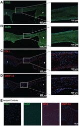
- Figure 8. Composition of tissue bridge formed by migratory cells. Immunohistochemical staining of tissue sections using antibodies reactive with (A) COL2, (B) ACAN, (C) COL1, or (D) MMP-13. (E) Isotype negative controls for COL2, ACAN, COL1, and MMP-13. *Bioengineered cartilage. #Native cartilage. The white box indicates the site of the magnified image of the tissue bridge. N = 3 biological replicates, (1 technical replicate). ACAN, aggrecan; COL, collagen; MMP, matrix metalloprotease.
