Explore
 Validate
 Learn
Learn Western blot
Antibody data
- Antibody Data
- Antigen structure
- References [6]
- Comments [0]
- Validations
- Western blot [2]
- Immunocytochemistry [1]
Submit
Validation data
Reference
Comment
Report error
- Product number
- PA1-1745 - Provider product page

- Provider
- Invitrogen Antibodies
- Product name
- Anti-Aggrecan G3 Polyclonal Antibody
- Antibody type
- Polyclonal
- Antigen
- Synthetic peptide
- Description
- PA1-1745 detects human, rat, mouse, bovine, and canine aggrecan G3. PA1-1745 has been successfully used in Western blot procedures. By Western blot, this antibody detects an ~350 kDa protein representing the aggrecan G3 domain on the full length core. The PA1-1745 immunogen is a synthetic peptide corresponding to residues C D(2277) G H P M Q F E N W R P N Q P D N(2293) of human aggrecan.. This antibody is also referred to as anti-G3.
- Reactivity
- Human, Mouse, Rat, Bovine, Canine
- Host
- Rabbit
- Isotype
- IgG
- Vial size
- 100 µg
- Concentration
- 1 mg/ml
- Storage
- -20° C, Avoid Freeze/Thaw Cycles
Submitted references Biochemical and atomic force microscopic characterization of salmon nasal cartilage proteoglycan.
MMP proteolysis of the human extracellular matrix protein aggrecan is mainly a process of normal turnover.
Aggrecanase cleavage in juvenile idiopathic arthritis patients is minimally detected in the aggrecan interglobular domain but robust at the aggrecan C-terminus.
Identification of proteoglycan from salmon nasal cartilage.
Calpain is involved in C-terminal truncation of human aggrecan.
A comparison of different purification methods of aggrecan fragments from human articular cartilage and synovial fluid.
Kakizaki I, Mineta T, Sasaki M, Tatara Y, Makino E, Kato Y
Carbohydrate polymers 2014 Mar 15;103:538-49
Carbohydrate polymers 2014 Mar 15;103:538-49
MMP proteolysis of the human extracellular matrix protein aggrecan is mainly a process of normal turnover.
Struglics A, Hansson M
The Biochemical journal 2012 Sep 1;446(2):213-23
The Biochemical journal 2012 Sep 1;446(2):213-23
Aggrecanase cleavage in juvenile idiopathic arthritis patients is minimally detected in the aggrecan interglobular domain but robust at the aggrecan C-terminus.
Struglics A, Lohmander LS, Last K, Akikusa J, Allen R, Fosang AJ
Arthritis and rheumatism 2012 Dec;64(12):4151-61; author reply 4162-3
Arthritis and rheumatism 2012 Dec;64(12):4151-61; author reply 4162-3
Identification of proteoglycan from salmon nasal cartilage.
Kakizaki I, Tatara Y, Majima M, Kato Y, Endo M
Archives of biochemistry and biophysics 2011 Feb 1;506(1):58-65
Archives of biochemistry and biophysics 2011 Feb 1;506(1):58-65
Calpain is involved in C-terminal truncation of human aggrecan.
Struglics A, Hansson M
The Biochemical journal 2010 Sep 15;430(3):531-8
The Biochemical journal 2010 Sep 15;430(3):531-8
A comparison of different purification methods of aggrecan fragments from human articular cartilage and synovial fluid.
Struglics A, Larsson S
Matrix biology : journal of the International Society for Matrix Biology 2010 Jan;29(1):74-83
Matrix biology : journal of the International Society for Matrix Biology 2010 Jan;29(1):74-83
No comments: Submit comment
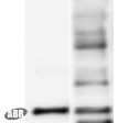
Supportive validation
- Submitted by
- Invitrogen Antibodies (provider)
- Main image

- Experimental details
- Figure 1 illustrates Western blot detection of mouse chondrocyte cells using PA1-1745. The specificity of PA1-1745 is demonstrated in lane 1. Lane 1: mouse chondrocyte Lane 2: a lane of human cartilage proteoglycan blotted with anti-aggrecan G1 domain (general)
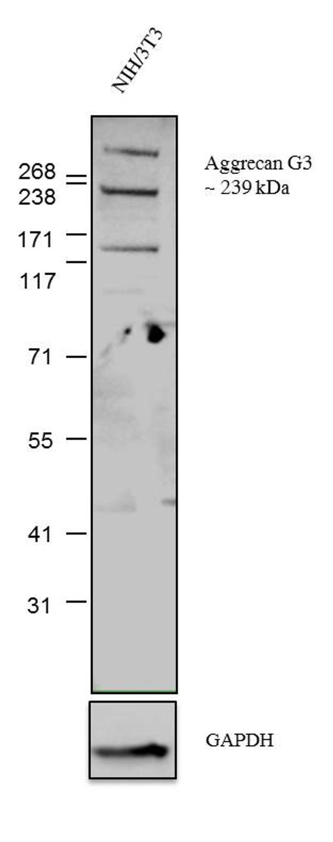
- Submitted by
- Invitrogen Antibodies (provider)
- Main image
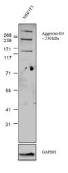
- Experimental details
- Western blot analysis was performed on whole cell extracts (30 µg lysate) of NIH/3T3 (Lane 1). The blots were probed with Anti-Aggrecan G3 alpha Rabbit Polyclonal Antibody (Product # PA1-1745, 1-3 µg/ml) and detected by chemiluminescence using Goat anti-Rabbit IgG (H+L) Superclonal™ Secondary Antibody, HRP conj µgate (Product # A27036, 0.4 µg/ml, 1:2500 dilution). A 239 kDa band corresponding to Aggrecan G3 was observed in the cell line tested. A lower band of 137 kDa was observed showing the proteolytic fragment of Aggrecan G3 and a higher band of ~300 kDa is a core protein which has around 100 GAG (glycosaminoglycans) chains attached to it. Known quantity of protein samples were electrophoresed using Novex® NuPAGE® 10% Bis-Tris gel (Product # NP0302BOX), XCell SureLock™ Electrophoresis System (Product # EI0002) and HiMark™ Pre-stained Protein Standard (Product # LC5699). Resolved proteins were then transferred onto a nitrocellulose membrane with overnight wet transfer System. The membrane was probed with the relevant primary and secondary Antibody following blocking with 5% skimmed milk. Chemiluminescent detection was performed using Novex® ECL Chemiluminescent Substrate Reagent Kit (Product # WP20005).
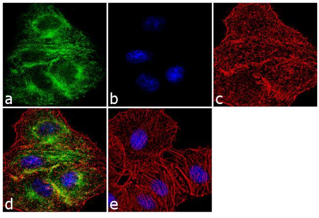
Supportive validation
- Submitted by
- Invitrogen Antibodies (provider)
- Main image
- Experimental details
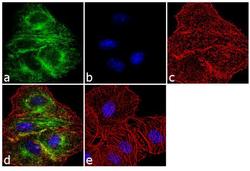
- Immunofluorescence analysis of Aggrecan G3 was performed using 70% confluent log phase A549 cells. The cells were fixed with 4% paraformaldehyde for 10 minutes, permeabilized with 0.1% Triton™ X-100 for 10 minutes, and blocked with 1% BSA for 1 hour at room temperature. The cells were labeled with Aggrecan G3 Rabbit Polyclonal Antibody (Product # PA1-1745) at 2 µg/ml in 0.1% BSA and incubated for 3 hours at room temperature and then labeled with Goat anti-Rabbit IgG (H+L) Superclonal™ Secondary Antibody, Alexa Fluor® 488 conj µgate (Product # A27034) at a dilution of 1:2000 for 45 minutes at room temperature (Panel a: green). Nuclei (Panel b: blue) were stained with SlowFade® Gold Antifade Mountant with DAPI (Product # S36938). F-actin (Panel c: red) was stained with Alexa Fluor® 555 Rhodamine Phalloidin (Product # R415, 1:300). Panel d represents the merged image showing localization in the extracellular matrix. Panel e shows the no primary antibody control. The images were captured at 60X magnification.