 Explore
 Validate
 Learn
Learn Western blot
Antibody data
- Antibody Data
- Antigen structure
- References [7]
- Comments [0]
- Validations
- Western blot [3]
- Immunohistochemistry [4]
Submit
Validation data
Reference
Comment
Report error
- Product number
- MA5-11276 - Provider product page

- Provider
- Invitrogen Antibodies
- Product name
- E2F4 Monoclonal Antibody (4E2F04 (WUF10))
- Antibody type
- Monoclonal
- Antigen
- Recombinant full-length protein
- Description
- MA5-11276 targets E2F-4 Transcription Factor in GS, IF, IHC (P), and WB applications and shows reactivity with Human and Rat samples.
- Antibody clone number
- 4E2F04 (WUF10)
- Concentration
- 0.2 mg/mL
Submitted references Anti-apoptotic function of the E2F transcription factor 4 (E2F4)/p130, a member of retinoblastoma gene family in cardiac myocytes.
Effects of isotretinoin on spermatogenesis of rats.
Classical Hodgkin lymphoma is characterized by high constitutive expression of activating transcription factor 3 (ATF3), which promotes viability of Hodgkin/Reed-Sternberg cells.
High-resolution analysis of 16q22.1 in breast carcinoma using DNA amplifiable probes (multiplex amplifiable probe hybridization technique) and immunohistochemistry.
p107 inhibits G1 to S phase progression by down-regulating expression of the F-box protein Skp2.
The adenovirus E4-6/7 protein directs nuclear localization of E2F-4 via an arginine-rich motif.
Expression of E2F-4 gene in colorectal adenocarcinoma and corresponding covering mucosa: an immunohistochemistry, image analysis, and immunoblot study.
Dingar D, Konecny F, Zou J, Sun X, von Harsdorf R
Journal of molecular and cellular cardiology 2012 Dec;53(6):820-8
Journal of molecular and cellular cardiology 2012 Dec;53(6):820-8
Effects of isotretinoin on spermatogenesis of rats.
Gencoglan G, Tosun M
Cutaneous and ocular toxicology 2011 Mar;30(1):55-60
Cutaneous and ocular toxicology 2011 Mar;30(1):55-60
Classical Hodgkin lymphoma is characterized by high constitutive expression of activating transcription factor 3 (ATF3), which promotes viability of Hodgkin/Reed-Sternberg cells.
Janz M, Hummel M, Truss M, Wollert-Wulf B, Mathas S, Jöhrens K, Hagemeier C, Bommert K, Stein H, Dörken B, Bargou RC
Blood 2006 Mar 15;107(6):2536-9
Blood 2006 Mar 15;107(6):2536-9
High-resolution analysis of 16q22.1 in breast carcinoma using DNA amplifiable probes (multiplex amplifiable probe hybridization technique) and immunohistochemistry.
Rakha EA, Armour JA, Pinder SE, Paish CE, Ellis IO
International journal of cancer 2005 May 1;114(5):720-9
International journal of cancer 2005 May 1;114(5):720-9
p107 inhibits G1 to S phase progression by down-regulating expression of the F-box protein Skp2.
Rodier G, Makris C, Coulombe P, Scime A, Nakayama K, Nakayama KI, Meloche S
The Journal of cell biology 2005 Jan 3;168(1):55-66
The Journal of cell biology 2005 Jan 3;168(1):55-66
The adenovirus E4-6/7 protein directs nuclear localization of E2F-4 via an arginine-rich motif.
Schaley JE, Polonskaia M, Hearing P
Journal of virology 2005 Feb;79(4):2301-8
Journal of virology 2005 Feb;79(4):2301-8
Expression of E2F-4 gene in colorectal adenocarcinoma and corresponding covering mucosa: an immunohistochemistry, image analysis, and immunoblot study.
Mady HH, Hasso S, Melhem MF
Applied immunohistochemistry & molecular morphology : AIMM 2002 Sep;10(3):225-30
Applied immunohistochemistry & molecular morphology : AIMM 2002 Sep;10(3):225-30
No comments: Submit comment
Supportive validation
- Submitted by
- Invitrogen Antibodies (provider)
- Main image

- Experimental details
- Western blot of E2F-4 Transcription Factor using E2F-4 Transcription Factor Monoclonal Antibody (Product # MA5-11276) on Raji Cells.

- Submitted by
- Invitrogen Antibodies (provider)
- Main image

- Experimental details
- Western blot analysis was performed on modified whole cell extracts (1% SDS) (30 µg lysate) of HCT 116 (Lane 1), SW-480 (Lane 2), HT-29 (Lane 3), COLO 205 (Lane 4), MDA-MB-231 (Lane 5) and Raji (Lane 6). The blot was probed with Anti-E2F4 Monoclonal Antibody (Product # MA5-11276, 2 µg/ml) and detected by chemiluminescence using Goat anti-Mouse IgG (H+L) Superclonal™ Secondary Antibody, HRP conjugate (Product # A28177, 0.25 µg/ml, 1:4000 dilution). A 50 kDa band corresponding to E2F4 was observed across the cell lines tested.

- Submitted by
- Invitrogen Antibodies (provider)
- Main image

- Experimental details
- Knockdown of E2F4 was achieved by transfecting HCT 116 with E2F4 specific siRNAs (Silencer® select Product # s4414). Western blot analysis (Fig. a) was performed using modified whole cell extracts (1% SDS) from the E2F4 knockdown cells (lane 3), non-specific scrambled siRNA transfected cells (lane 2) and untransfected cells (lane 1). The blots were probed with E2F4 Monoclonal Antibody (4E2F04 (WUF10)) (Product # MA5-11276, 2 µg/ml) and Goat anti-Mouse IgG (H+L) Superclonal™ Secondary Antibody, HRP conjugate (Product # A28177, 0.25 µg/ml, 1:4000 dilution). Densitometric analysis of this western blot is shown in histogram (Fig. b). Loss of signal upon siRNA mediated knock down confirms that antibody is specific to E2F4.

Supportive validation
- Submitted by
- Invitrogen Antibodies (provider)
- Main image
- Experimental details
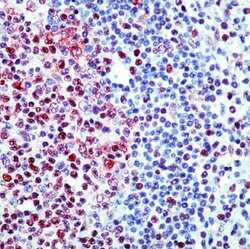
- Formalin-fixed, paraffin-embedded human tonsil stained with E2F-4 antibody using peroxidase-conjugate and AEC chromogen. Note nuclear staining of cells.

- Submitted by
- Invitrogen Antibodies (provider)
- Main image
- Experimental details
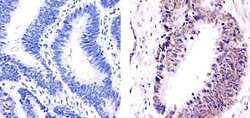
- Immunohistochemistry analysis of E2F-4 Transcription Factor showing staining in the nucleus of paraffin-embedded human colon adenocarcinoma (right) compared to a negative control without primary antibody (left). To expose target proteins, antigen retrieval was performed using 10mM sodium citrate (pH 6.0), microwaved for 8-15 min. Following antigen retrieval, tissues were blocked in 3% H2O2-methanol for 15 min at room temperature, washed with ddH2O and PBS, and then probed with a E2F-4 Transcription Factor Mouse Monoclonal Antibody (Product # MA5-11276) diluted in 3% BSA-PBS at a dilution of 1:20 for 1 hour at 37°C in a humidified chamber. Tissues were washed extensively in PBST and detection was performed using an HRP-conjugated secondary antibody followed by colorimetric detection using a DAB kit. Tissues were counterstained with hematoxylin and dehydrated with ethanol and xylene to prep for mounting.

- Submitted by
- Invitrogen Antibodies (provider)
- Main image
- Experimental details
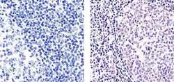
- Immunohistochemistry analysis of E2F-4 Transcription Factor showing staining in the nucleus of paraffin-embedded human tonsil tissue (right) compared to a negative control without primary antibody (left). To expose target proteins, antigen retrieval was performed using 10mM sodium citrate (pH 6.0), microwaved for 8-15 min. Following antigen retrieval, tissues were blocked in 3% H2O2-methanol for 15 min at room temperature, washed with ddH2O and PBS, and then probed with a E2F-4 Transcription Factor Mouse Monoclonal Antibody (Product # MA5-11276) diluted in 3% BSA-PBS at a dilution of 1:20 for 1 hour at 37°C in a humidified chamber. Tissues were washed extensively in PBST and detection was performed using an HRP-conjugated secondary antibody followed by colorimetric detection using a DAB kit. Tissues were counterstained with hematoxylin and dehydrated with ethanol and xylene to prep for mounting.

- Submitted by
- Invitrogen Antibodies (provider)
- Main image
- Experimental details
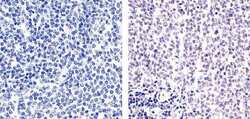
- Immunohistochemistry analysis of E2F-4 Transcription Factor showing staining in the nucleus of paraffin-embedded mouse spleen tissue (right) compared to a negative control without primary antibody (left). To expose target proteins, antigen retrieval was performed using 10mM sodium citrate (pH 6.0), microwaved for 8-15 min. Following antigen retrieval, tissues were blocked in 3% H2O2-methanol for 15 min at room temperature, washed with ddH2O and PBS, and then probed with a E2F-4 Transcription Factor Mouse Monoclonal Antibody (Product # MA5-11276) diluted in 3% BSA-PBS at a dilution of 1:20 for 1 hour at 37°C in a humidified chamber. Tissues were washed extensively in PBST and detection was performed using an HRP-conjugated secondary antibody followed by colorimetric detection using a DAB kit. Tissues were counterstained with hematoxylin and dehydrated with ethanol and xylene to prep for mounting.
