 Explore
 Validate
 Learn
Learn Western blot
 ELISA
ELISAAntibody data
- Antibody Data
- Antigen structure
- References [12]
- Comments [0]
- Validations
- Western blot [1]
- Immunohistochemistry [2]
- Flow cytometry [1]
Submit
Validation data
Reference
Comment
Report error
- Product number
- 16226-1-AP - Provider product page

- Provider
- Proteintech Group
- Proper citation
- Proteintech Cat#16226-1-AP, RRID:AB_2252090
- Product name
- PDE4A antibody
- Antibody type
- Polyclonal
- Description
- KD/KO validated PDE4A antibody (Cat. #16226-1-AP) is a rabbit polyclonal antibody that shows reactivity with human, mouse, rat and has been validated for the following applications: FC, IHC, IP, WB,ELISA.
- Reactivity
- Human, Mouse, Rat
- Host
- Rabbit
- Conjugate
- Unconjugated
- Isotype
- IgG
- Vial size
- 20ul, 150ul
Submitted references Inhibition of phosphodiesterase-4 by rolipram alleviates anxiety-like behavior in mice with PTSD by modulating neuroinflammation and synaptic plasticity via cAMP signaling.
Ginsenoside Rg1 attenuates Aβ(1-42)-induced microglial cell apoptosis and inflammation in Alzheimer's disease via the GATA4/PDE4A/PI3K/AKT axis.
Upregulation of Phosphodiesterase 7A Contributes to Concurrent Pain and Depression via Inhibition of cAMP-PKA-CREB-BDNF Signaling and Neuroinflammation in the Hippocampus of Mice.
Potential PDE4A inhibition-mediated neuroprotective effects of psoralidin.
Rolipram Ameliorates Memory Deficits and Depression-Like Behavior in APP/PS1/tau Triple Transgenic Mice: Involvement of Neuroinflammation and Apoptosis via cAMP Signaling.
Involvement of 3',5'-cyclic inosine monophosphate in cystathionine γ-lyase-dependent regulation of the vascular tone.
Phosphodiesterases Expression during Murine Cardiac Development.
RGS10 physically and functionally interacts with STIM2 and requires store-operated calcium entry to regulate pro-inflammatory gene expression in microglia.
Inhibition of PDE4/PDE4B improves renal function and ameliorates inflammation in cisplatin-induced acute kidney injury.
DC591017, a phosphodiesterase-4 (PDE4) inhibitor with robust anti-inflammation through regulating PKA-CREB signaling.
Apremilast Normalizes Gene Expression of Inflammatory Mediators in Human Keratinocytes and Reduces Antigen-Induced Atopic Dermatitis in Mice.
Phosphodiesterase 4 in inflammatory diseases: Effects of apremilast in psoriatic blood and in dermal myofibroblasts through the PDE4/CD271 complex.
Lin Y, Ma S, Sun R, Ruan M, Zhang F, Zhang HT
International immunopharmacology 2025 Sep 23;162:115116
International immunopharmacology 2025 Sep 23;162:115116
Ginsenoside Rg1 attenuates Aβ(1-42)-induced microglial cell apoptosis and inflammation in Alzheimer's disease via the GATA4/PDE4A/PI3K/AKT axis.
Fang H, Tian H, Liu J, Peng T, Wang D
Neuroscience 2025 Jan 26;565:377-385
Neuroscience 2025 Jan 26;565:377-385
Upregulation of Phosphodiesterase 7A Contributes to Concurrent Pain and Depression via Inhibition of cAMP-PKA-CREB-BDNF Signaling and Neuroinflammation in the Hippocampus of Mice.
Chen SC, Chen YH, Song Y, Zong SH, Wu MX, Wang W, Wang H, Zhang F, Zhou YM, Yu HY, Zhang HT, Zhang FF
The international journal of neuropsychopharmacology 2024 Oct 1;27(10)
The international journal of neuropsychopharmacology 2024 Oct 1;27(10)
Potential PDE4A inhibition-mediated neuroprotective effects of psoralidin.
Uzunhisarcıklı E
European review for medical and pharmacological sciences 2024 Nov;28(21):4546-4552
European review for medical and pharmacological sciences 2024 Nov;28(21):4546-4552
Rolipram Ameliorates Memory Deficits and Depression-Like Behavior in APP/PS1/tau Triple Transgenic Mice: Involvement of Neuroinflammation and Apoptosis via cAMP Signaling.
Cong YF, Liu FW, Xu L, Song SS, Shen XR, Liu D, Hou XQ, Zhang HT
The international journal of neuropsychopharmacology 2023 Sep 25;26(9):585-598
The international journal of neuropsychopharmacology 2023 Sep 25;26(9):585-598
Involvement of 3',5'-cyclic inosine monophosphate in cystathionine γ-lyase-dependent regulation of the vascular tone.
Mitidieri E, Vellecco V, Brancaleone V, Vanacore D, Manzo OL, Martin E, Sharina I, Krutsenko Y, Monti MC, Morretta E, Papapetropoulos A, Caliendo G, Frecentese F, Cirino G, Sorrentino R, d'Emmanuele di Villa Bianca R, Bucci M
British journal of pharmacology 2021 Sep;178(18):3765-3782
British journal of pharmacology 2021 Sep;178(18):3765-3782
Phosphodiesterases Expression during Murine Cardiac Development.
Carvalho TMDCS, Cardarelli S, Giorgi M, Lenzi A, Isidori AM, Naro F
International journal of molecular sciences 2021 Mar 5;22(5)
International journal of molecular sciences 2021 Mar 5;22(5)
RGS10 physically and functionally interacts with STIM2 and requires store-operated calcium entry to regulate pro-inflammatory gene expression in microglia.
Wendimu MY, Alqinyah M, Vella S, Dean P, Almutairi F, Davila-Rivera R, Rayatpisheh S, Wohlschlegel J, Moreno S, Hooks SB
Cellular signalling 2021 Jul;83:109974
Cellular signalling 2021 Jul;83:109974
Inhibition of PDE4/PDE4B improves renal function and ameliorates inflammation in cisplatin-induced acute kidney injury.
Xu M, Yu X, Meng X, Huang S, Zhang Y, Zhang A, Jia Z
American journal of physiology. Renal physiology 2020 Mar 1;318(3):F576-F588
American journal of physiology. Renal physiology 2020 Mar 1;318(3):F576-F588
DC591017, a phosphodiesterase-4 (PDE4) inhibitor with robust anti-inflammation through regulating PKA-CREB signaling.
Li H, Li J, Zhang X, Feng C, Fan C, Yang X, Zhang R, Zhu F, Zhou Y, Xu Y, Liu H, Tang W
Biochemical pharmacology 2020 Jul;177:113958
Biochemical pharmacology 2020 Jul;177:113958
Apremilast Normalizes Gene Expression of Inflammatory Mediators in Human Keratinocytes and Reduces Antigen-Induced Atopic Dermatitis in Mice.
Schafer PH, Adams M, Horan G, Truzzi F, Marconi A, Pincelli C
Drugs in R&D 2019 Dec;19(4):329-338
Drugs in R&D 2019 Dec;19(4):329-338
Phosphodiesterase 4 in inflammatory diseases: Effects of apremilast in psoriatic blood and in dermal myofibroblasts through the PDE4/CD271 complex.
Schafer PH, Truzzi F, Parton A, Wu L, Kosek J, Zhang LH, Horan G, Saltari A, Quadri M, Lotti R, Marconi A, Pincelli C
Cellular signalling 2016 Jul;28(7):753-63
Cellular signalling 2016 Jul;28(7):753-63
No comments: Submit comment
Supportive validation
- Submitted by
- Proteintech Group (provider)
- Main image
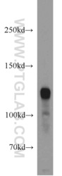
- Experimental details
- human brain tissue were subjected to SDS PAGE followed by western blot with 16226-1-AP(PDE4A antibody) at dilution of 1:1000
- Sample type
- tissue

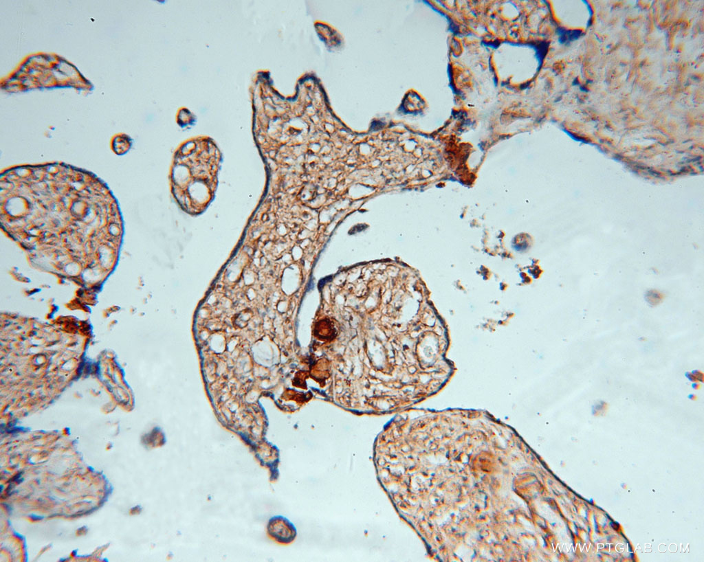
Supportive validation
- Submitted by
- Proteintech Group (provider)
- Main image

- Experimental details
- Immunohistochemical of paraffin-embedded human placenta using 16226-1-AP(PDE4A antibody) at dilution of 1:50 (under 10x lens)
- Sample type
- tissue
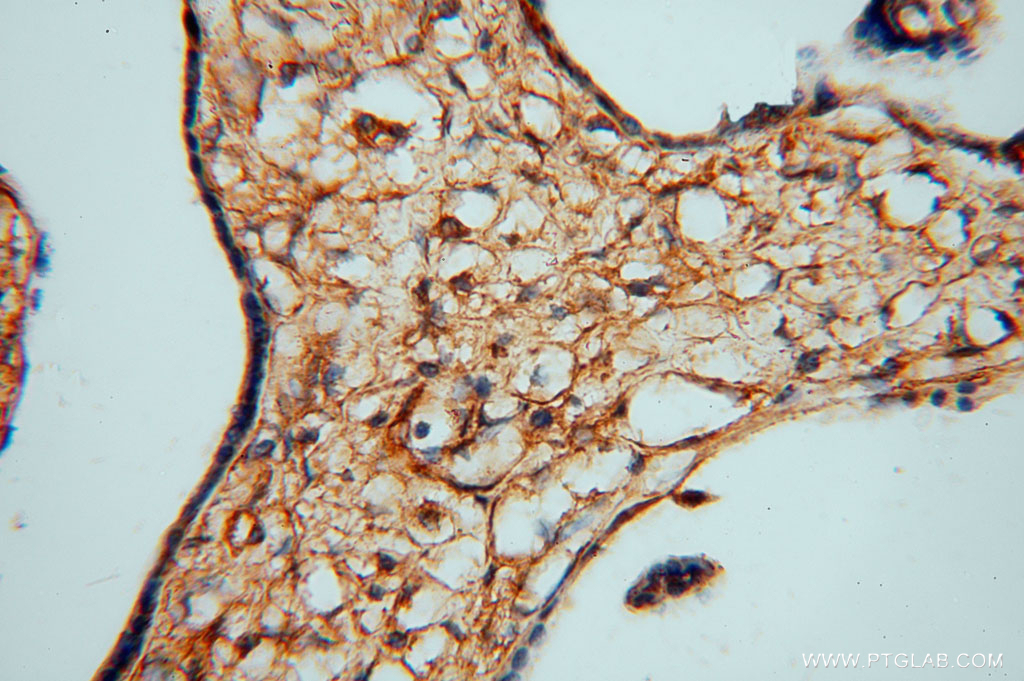
- Submitted by
- Proteintech Group (provider)
- Main image

- Experimental details
- Immunohistochemical of paraffin-embedded human placenta using 16226-1-AP(PDE4A antibody) at dilution of 1:50 (under 40x lens)
- Sample type
- tissue
Supportive validation
- Submitted by
- Proteintech Group (provider)
- Main image
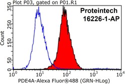
- Experimental details
- The PDE4A antibody from Proteintech is a rabbit polyclonal antibody to a recombinant protein of human PDE4A. This antibody recognizes human,mouse,rat antigen. The PDE4A antibody has been validated for the following applications: ELISA, WB, IHC, FC analysis.
