Explore
 Validate
 Learn
Learn Western blot
 Immunocytochemistry
Antibody data
- Antibody Data
- Antigen structure
- References [0]
- Comments [0]
- Validations
- Western blot [1]
- Immunohistochemistry [8]
- Flow cytometry [1]
Submit
Validation data
Reference
Comment
Report error
- Product number
- NBP2-02559 - Provider product page

- Provider
- Novus Biologicals
- Product name
- Mouse Monoclonal Phosphodiesterase 4A/PDE4A Antibody
- Antibody type
- Monoclonal
- Description
- Affinity purified.
- Reactivity
- Human
- Host
- Mouse
- Isotype
- IgG
- Vial size
- 0.1 ml
- Concentration
- 0.92 mg/ml
- Storage
- Store at -20C. Avoid freeze-thaw cycles.
No comments: Submit comment
Supportive validation
- Submitted by
- Novus Biologicals (provider)
- Main image
- Experimental details
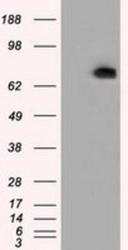
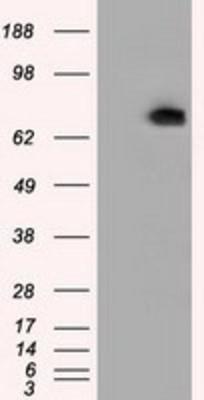
- Western Blot: Phosphodiesterase 4A/PDE4A Antibody (1C8) [NBP2-02559] - HEK293T cells were transfected with the pCMV6-ENTRY control (Left lane) or pCMV6-ENTRY PDE4A (Right lane) cDNA for 48 hrs and lysed. Equivalent amounts of cell lysates (5 ug per lane) were separated by SDS-PAGE and immunoblotted with anti-PDE4A.
Supportive validation
- Submitted by
- Novus Biologicals (provider)
- Main image
- Experimental details
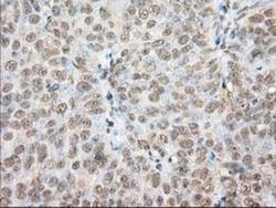
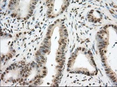
- Immunohistochemistry-Paraffin: Phosphodiesterase 4A/PDE4A Antibody (1C8) [NBP2-02559] - Staining of paraffin-embedded Adenocarcinoma of Human ovary tissue using anti-PDE4A mouse monoclonal antibody.

- Submitted by
- Novus Biologicals (provider)
- Main image

- Experimental details
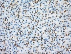
- Immunohistochemistry-Paraffin: Phosphodiesterase 4A/PDE4A Antibody (1C8) [NBP2-02559] - Staining of paraffin-embedded Adenocarcinoma of Human colon tissue using anti-PDE4A mouse monoclonal antibody.
- Submitted by
- Novus Biologicals (provider)
- Main image
- Experimental details
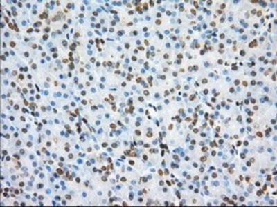
- Immunohistochemistry-Paraffin: Phosphodiesterase 4A/PDE4A Antibody (1C8) [NBP2-02559] - Staining of paraffin-embedded Human pancreas tissue using anti-PDE4A mouse monoclonal antibody.
- Submitted by
- Novus Biologicals (provider)
- Main image

- Experimental details
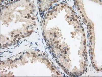
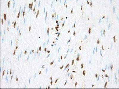
- Immunohistochemistry-Paraffin: Phosphodiesterase 4A/PDE4A Antibody (1C8) [NBP2-02559] - Staining of paraffin-embedded Human prostate tissue using anti-PDE4A mouse monoclonal antibody.
- Submitted by
- Novus Biologicals (provider)
- Main image
- Experimental details
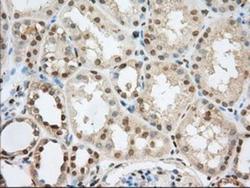
- Immunohistochemistry-Paraffin: Phosphodiesterase 4A/PDE4A Antibody (1C8) [NBP2-02559] - Staining of paraffin-embedded Human Kidney tissue using anti-PDE4A mouse monoclonal antibody.

- Submitted by
- Novus Biologicals (provider)
- Main image
- Experimental details
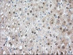
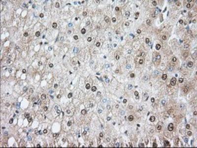
- Immunohistochemistry-Paraffin: Phosphodiesterase 4A/PDE4A Antibody (1C8) [NBP2-02559] - Staining of paraffin-embedded Human liver tissue using anti-PDE4A mouse monoclonal antibody.
- Submitted by
- Novus Biologicals (provider)
- Main image
- Experimental details
- Immunohistochemistry-Paraffin: Phosphodiesterase 4A/PDE4A Antibody (1C8) [NBP2-02559] - Staining of paraffin-embedded Human colon tissue using anti-PDE4A mouse monoclonal antibody.
- Submitted by
- Novus Biologicals (provider)
- Main image
- Experimental details
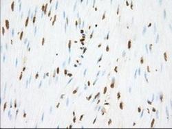
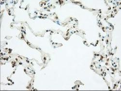
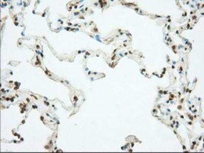
- Immunohistochemistry-Paraffin: Phosphodiesterase 4A/PDE4A Antibody (1C8) [NBP2-02559] - Staining of paraffin-embedded Human lung tissue using anti-PDE4A mouse monoclonal antibody.
Supportive validation
- Submitted by
- Novus Biologicals (provider)
- Main image
- Experimental details
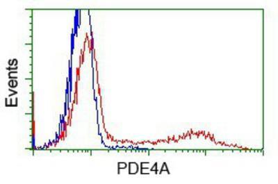
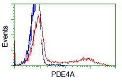
- Flow Cytometry: Phosphodiesterase 4A/PDE4A Antibody (1C8) [NBP2-02559] - HEK293T cells transfected with either overexpression plasmid (Red) or empty vector control plasmid (Blue) were immunostained by anti-PDE4A antibody, and then analyzed by flow cytometry.