Explore
 Validate
 Learn
LearnHPA052818
antibody from Atlas Antibodies
Targeting: MED1
CRSP1, CRSP200, DRIP230, PBP, PPARBP, PPARGBP, RB18A, TRAP220, TRIP2
 Immunocytochemistry
Antibody data
- Antibody Data
- Antigen structure
- References [2]
- Comments [0]
- Validations
- Immunocytochemistry [1]
- Chromatin Immunoprecipitation [1]
Submit
Validation data
Reference
Comment
Report error
- Product number
- HPA052818 - Provider product page

- Provider
- Atlas Antibodies
- Proper citation
- Atlas Antibodies Cat#HPA052818, RRID:AB_2681962
- Product name
- Anti-MED1
- Antibody type
- Polyclonal
- Description
- Polyclonal Antibody against Human MED1, Gene description: mediator complex subunit 1, Alternative Gene Names: CRSP1, CRSP200, DRIP230, PBP, PPARBP, PPARGBP, RB18A, TRAP220, TRIP2, Validated applications: ICC, ChIP, Uniprot ID: Q15648, Storage: Store at +4°C for short term storage. Long time storage is recommended at -20°C.
- Reactivity
- Human
- Host
- Rabbit
- Conjugate
- Unconjugated
- Isotype
- IgG
- Vial size
- 100 µl
- Concentration
- 0.1 mg/ml
- Storage
- Store at +4°C for short term storage. Long time storage is recommended at -20°C.
- Handling
- The antibody solution should be gently mixed before use.
Submitted references Notch1 Phase Separation Coupled Percolation facilitates target gene expression and enhancer looping.
Regulated splicing of large exons is linked to phase‐separation of vertebrate transcription factors
Foran G, Hallam RD, Megaly M, Turgambayeva A, Antfolk D, Li Y, Luca VC, Necakov A
bioRxiv : the preprint server for biology 2024 Aug 1;
bioRxiv : the preprint server for biology 2024 Aug 1;
Regulated splicing of large exons is linked to phase‐separation of vertebrate transcription factors
Kawachi T, Masuda A, Yamashita Y, Takeda J, Ohkawara B, Ito M, Ohno K
The EMBO Journal 2021;40(22)
The EMBO Journal 2021;40(22)
No comments: Submit comment
Supportive validation
- Submitted by
- Atlas Antibodies (provider)
- Main image
- Experimental details
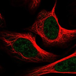
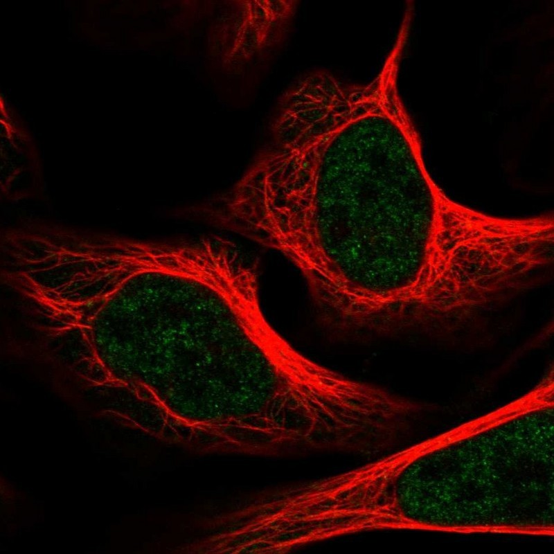
- Immunofluorescent staining of human cell line U-2 OS shows localization to nucleoplasm.
- Sample type
- Human
Supportive validation
- Submitted by
- Atlas Antibodies (provider)
- Main image

- Experimental details
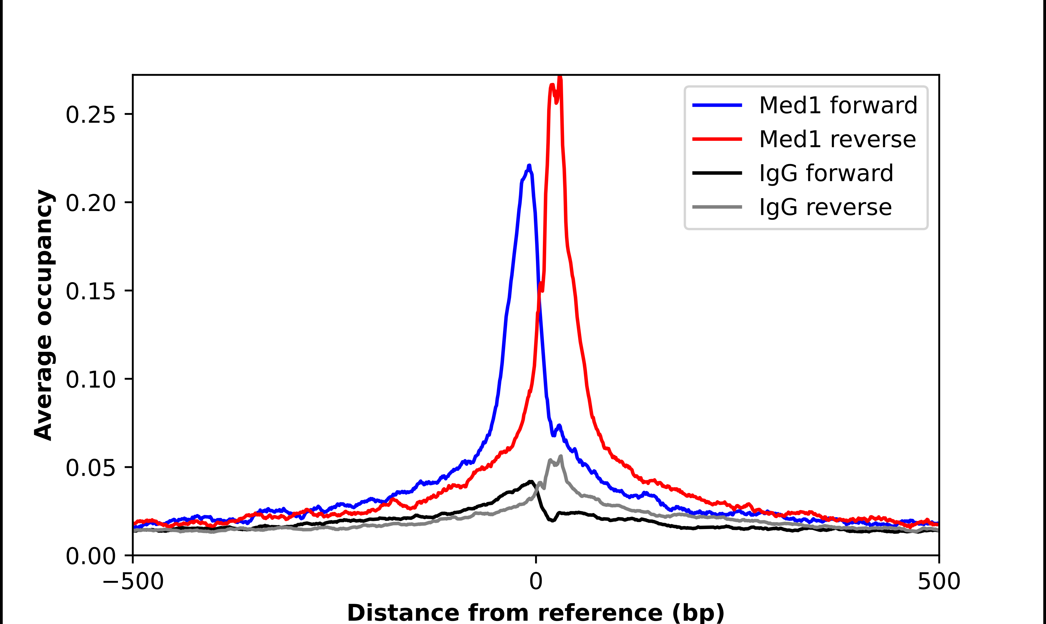
- ChIP-Exo-Seq composite graph for Anti-MED1 (HPA052818, Lot 000011173) tested in K562 cells. Strand-specific reads (blue: forward, red: reverse) and IgG controls (black: forward, grey: reverse) are plotted against the distance from a composite set of reference binding sites. The antibody exhibits robust target enrichment compared to a non-specific IgG control and precisely reveals its structural organization around the binding site. Data generated by Prof. B. F. Pugh´s Lab at Cornell University.