Explore
 Validate
 Learn
LearnMAB7650
antibody from Novus Biologicals
Targeting: TCF3
bHLHb21, E2A, E47, ITF1, MGC129647, MGC129648, p75, VDIR
 Western blot
Antibody data
- Antibody Data
- Antigen structure
- References [2]
- Comments [0]
- Validations
- Western blot [1]
- Immunohistochemistry [1]
Submit
Validation data
Reference
Comment
Report error
- Product number
- MAB7650 - Provider product page

- Provider
- Novus Biologicals
- Product name
- Rat Monoclonal TCF-3/E2A Antibody
- Antibody type
- Monoclonal
- Description
- Protein A or G purified from hybridoma culture supernatant. Detects mouse TCF-3/E2A in ELISAs. In direct ELISAs, no cross-reactivity with recombinant human TCF-3 is observed.
- Reactivity
- Mouse
- Host
- Rat
- Isotype
- IgG
- Vial size
- 100 ug
- Concentration
- LYOPH
- Storage
- Use a manual defrost freezer and avoid repeated freeze-thaw cycles. 12 months from date of receipt, -20 to -70 degreesC as supplied. 1 month, 2 to 8 degreesC under sterile conditions after reconstitution. 6 months, -20 to -70 degreesC under sterile conditions after reconstitution.
Submitted references Comparative genomics reveals multistep pathogenesis of E2A-PBX1 acute lymphoblastic leukemia.
De novo DNA Methyltransferases Dnmt3a and Dnmt3b regulate the onset of Igκ light chain rearrangement during early B-cell development.
Duque-Afonso J, Feng J, Scherer F, Lin CH, Wong SH, Wang Z, Iwasaki M, Cleary ML
The Journal of clinical investigation 2015 Sep;125(9):3667-80
The Journal of clinical investigation 2015 Sep;125(9):3667-80
De novo DNA Methyltransferases Dnmt3a and Dnmt3b regulate the onset of Igκ light chain rearrangement during early B-cell development.
Manoharan A, Du Roure C, Rolink AG, Matthias P
European journal of immunology 2015 Aug;45(8):2343-55
European journal of immunology 2015 Aug;45(8):2343-55
No comments: Submit comment
Supportive validation
- Submitted by
- Novus Biologicals (provider)
- Main image
- Experimental details
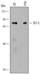
- Detection of Mouse TCF-3/E2A by Western Blot. Western blot shows lysates of D3 mouse embryonic stem cell line and P19 mouse embryonal carcinoma cell line. PVDF membrane was probed with 2 µg/mL of Rat Anti-Mouse TCF-3/E2A Monoclonal Antibody (Catalog # MAB7650) followed by HRP-conjugated Anti-Rat IgG Secondary Antibody (Catalog # HAF005). A specific band was detected for TCF-3/E2A at approximately 75 kDa (as indicated). This experiment was conducted under reducing conditions and using Immunoblot Buffer Group 1.

Supportive validation
- Submitted by
- Novus Biologicals (provider)
- Main image

- Experimental details
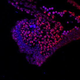
- TCF-3/E2A in Mouse Embryo. TCF-3/E2A was detected in immersion fixed frozen sections of E9.5 mouse embryo using Rat Anti-Mouse TCF-3/E2A Monoclonal Antibody (Catalog # MAB7650) at 10 µg/mL overnight at 4 °C. Tissue was stained using the NorthernLights™ 557-conjugated Anti-Rat IgG Secondary Antibody (red; Catalog # NL013) and counterstained with DAPI (blue). Specific staining was localized to branchial arch nuclei. View our protocol for Fluorescent IHC Staining of Frozen Tissue Sections.