 Explore
 Validate
 Learn
Learn Western blot
 Flow cytometry
Antibody data
- Antibody Data
- Antigen structure
- References [0]
- Comments [0]
- Validations
- Western blot [5]
- Immunocytochemistry [1]
Submit
Validation data
Reference
Comment
Report error
- Product number
- MA5-25351 - Provider product page

- Provider
- Invitrogen Antibodies
- Product name
- ERK5 Monoclonal Antibody (OTI3D4)
- Antibody type
- Monoclonal
- Antigen
- Recombinant full-length protein
- Reactivity
- Human
- Host
- Mouse
- Isotype
- IgG
- Antibody clone number
- OTI3D4
- Vial size
- 100 µL
- Concentration
- 0.35 mg/mL
- Storage
- -20° C, Avoid Freeze/Thaw Cycles
No comments: Submit comment
Supportive validation
- Submitted by
- Invitrogen Antibodies (provider)
- Main image

- Experimental details
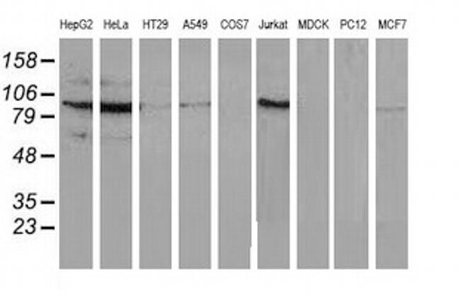
- Western blot analysis of MAPK7 in HepG2, HeLa, HT29, A549, COS7, Jurkat, MDCK, PC12, MCF7 cells using 35 µg per lane. Samples were probed with MAPK7 (Product # MA5-25351) monoclonal antibody.

- Submitted by
- Invitrogen Antibodies (provider)
- Main image

- Experimental details
- Western blot analysis of MAPK7 in HEK293T cells in untransfected (Left lane) and transfected (Right lane) samples using 5 µg per lane. The samples were separated by SDS-PAGE and probed with MAPK7 (Product # MA5-25351) monoclonal antibody.

- Submitted by
- Invitrogen Antibodies (provider)
- Main image

- Experimental details
- Western blot analysis of MAPK7 in HepG2, HeLa, HT29, A549, COS7, Jurkat, MDCK, PC12, MCF7 cells using 35 µg per lane. Samples were probed with MAPK7 (Product # MA5-25351) monoclonal antibody.
- Submitted by
- Invitrogen Antibodies (provider)
- Main image

- Experimental details
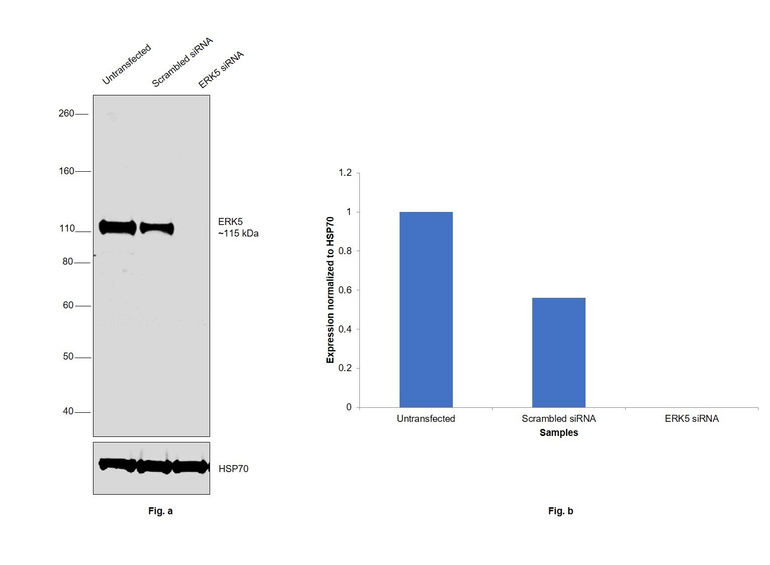
- Knockdown of Mitogen-activated protein kinase 7 was achieved by transfecting DU 145 with Mitogen-activated protein kinase 7 specific siRNAs (Silencer® select Product # s11149, s11151). Western blot analysis (Fig. a) was performed using Whole cell extracts from the Mitogen-activated protein kinase 7 knockdown cells (lane 3), non-targeting scrambled siRNA transfected cells (lane 2) and untransfected cells (lane 1). The blot was probed with ERK5 Monoclonal Antibody (OTI3D4) (Product # MA5-25351, 1:300 dilution ) and Goat anti-Mouse IgG (H+L) Superclonal™ Recombinant Secondary Antibody, HRP (Product # A28177, 1:6000 dilution). Densitometric analysis of this western blot is shown in histogram (Fig. b). Decrease in signal upon siRNA mediated knock down confirms that antibody is specific to Mitogen-activated protein kinase 7.
- Submitted by
- Invitrogen Antibodies (provider)
- Main image
- Experimental details
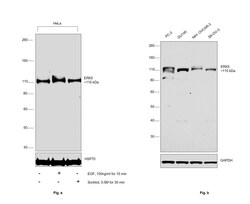
- Western blot was performed using Anti-ERK5 Monoclonal Antibody (OTI3D4) (Product # MA5-25351) and a 115 kDa band corresponding to Mitogen-activated protein kinase 7 was observed across cell lines tested. Upon EGF and sorbitol treatment in HeLa cells, there was a shift in the band size which corresponds to phosphorylated form of ERK5, since it is an autophosphorylated protein. Whole cell extracts (30 µg lysate) of (Fig.a) HeLa (Lane 1), HeLa serum starved for overnight and treated with EGF (100 ng/mL for 15 min) (Lane 2), HeLa treated with Sorbitol (0.5M for 30 min) (Lane 3) were electrophoresed using NuPAGE™ 3-8% Tris-Acetate Protein Gel (Product # EA0378BOX); (Fig.b) PC-3 (Lane 1), DU 145 (Lane 2), NIH:OVCAR-3 (Lane 3), SK-O-V3 (Lane 4). Resolved proteins were then transferred onto a Nitrocellulose membrane (Product # IB23001) by iBlot® 2 Dry Blotting System (Product # IB21001). The blot was probed with the primary antibody (1:300 dilution) and detected by chemiluminescence with Goat anti-Mouse IgG (H+L) Superclonal™ Recombinant Secondary Antibody, HRP (Product # A28177,1:6000 dilution) using the iBright FL 1000 (Product # A32752). Chemiluminescent detection was performed using SuperSignal™ West Dura Extended Duration Substrate (Product # 34076).

Supportive validation
- Submitted by
- Invitrogen Antibodies (provider)
- Main image
- Experimental details
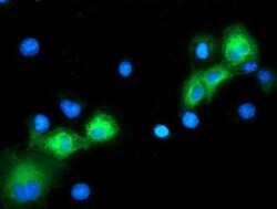
- Immunofluorescent analysis of MAPK7 in COS7 cells. Cells were transfected with a plasmid overexpressing MAPK7 and probed with a MAPK7 monoclonal antibody (Product # MA5-25351).
