 Explore
 Validate
 Learn
Learn37-7600
antibody from Invitrogen Antibodies
Targeting: LRP1
A2MR, APOER, APR, CD91, IGFBP-3R, IGFBP3R1, LRP, LRP1A
 Western blot
 Immunocytochemistry
Antibody data
- Antibody Data
- Antigen structure
- References [5]
- Comments [0]
- Validations
- Immunocytochemistry [2]
- Immunohistochemistry [1]
- Other assay [9]
Submit
Validation data
Reference
Comment
Report error
- Product number
- 37-7600 - Provider product page

- Provider
- Invitrogen Antibodies
- Product name
- LRP1 Monoclonal Antibody (A2MR-beta-1)
- Antibody type
- Monoclonal
- Antigen
- Synthetic peptide
- Description
- 37-7600 was used in IHC to successfully detect LRP-1 in mouse E18.5 lung section.
- Reactivity
- Human, Mouse, Rat
- Host
- Mouse
- Isotype
- IgG
- Antibody clone number
- A2MR-beta-1
- Vial size
- 100 μg
- Concentration
- 0.5 mg/mL
- Storage
- -20°C
Submitted references LRP-1 receptor combines EGFR signalling and eHsp90α autocrine to support constitutive breast cancer cell motility in absence of blood supply.
Human neural cell type-specific extracellular vesicle proteome defines disease-related molecules associated with activated astrocytes in Alzheimer's disease brain.
Breast Cancer MDA-MB-231 Cells Use Secreted Heat Shock Protein-90alpha (Hsp90α) to Survive a Hostile Hypoxic Environment.
Hsp90α and Hsp90β together operate a hypoxia and nutrient paucity stress-response mechanism during wound healing.
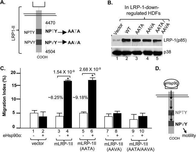
Extracellular heat shock protein 90 signals through subdomain II and the NPVY motif of LRP-1 receptor to Akt1 and Akt2: a circuit essential for promoting skin cell migration in vitro and wound healing in vivo.
Chang C, Tang X, Mosallaei D, Chen M, Woodley DT, Schönthal AH, Li W
Scientific reports 2022 Jul 14;12(1):12006
Scientific reports 2022 Jul 14;12(1):12006
Human neural cell type-specific extracellular vesicle proteome defines disease-related molecules associated with activated astrocytes in Alzheimer's disease brain.
You Y, Muraoka S, Jedrychowski MP, Hu J, McQuade AK, Young-Pearse T, Aslebagh R, Shaffer SA, Gygi SP, Blurton-Jones M, Poon WW, Ikezu T
Journal of extracellular vesicles 2022 Jan;11(1):e12183
Journal of extracellular vesicles 2022 Jan;11(1):e12183
Breast Cancer MDA-MB-231 Cells Use Secreted Heat Shock Protein-90alpha (Hsp90α) to Survive a Hostile Hypoxic Environment.
Dong H, Zou M, Bhatia A, Jayaprakash P, Hofman F, Ying Q, Chen M, Woodley DT, Li W
Scientific reports 2016 Feb 5;6:20605
Scientific reports 2016 Feb 5;6:20605
Hsp90α and Hsp90β together operate a hypoxia and nutrient paucity stress-response mechanism during wound healing.
Jayaprakash P, Dong H, Zou M, Bhatia A, O'Brien K, Chen M, Woodley DT, Li W
Journal of cell science 2015 Apr 15;128(8):1475-80
Journal of cell science 2015 Apr 15;128(8):1475-80
Extracellular heat shock protein 90 signals through subdomain II and the NPVY motif of LRP-1 receptor to Akt1 and Akt2: a circuit essential for promoting skin cell migration in vitro and wound healing in vivo.
Tsen F, Bhatia A, O'Brien K, Cheng CF, Chen M, Hay N, Stiles B, Woodley DT, Li W
Molecular and cellular biology 2013 Dec;33(24):4947-59
Molecular and cellular biology 2013 Dec;33(24):4947-59
No comments: Submit comment
Supportive validation
- Submitted by
- Invitrogen Antibodies (provider)
- Main image

- Experimental details
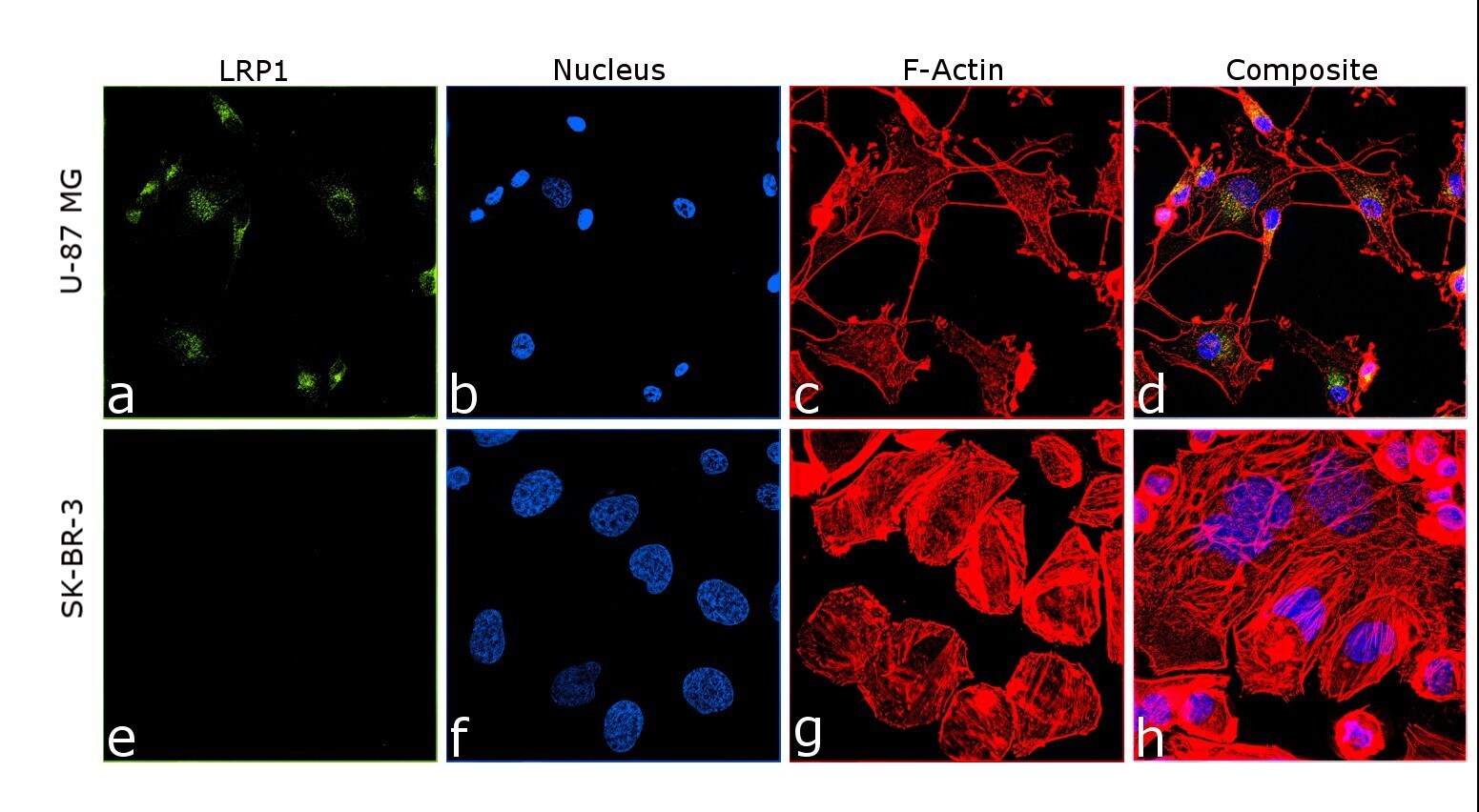
- Immunofluorescence analysis of LRP1 was performed using 70% confluent log phase U-87 MG and SK-BR-3 cells. The cells were fixed with 4% Paraformaldehyde for 10 minutes, permeabilized with 0.1% Triton™ X-100 for 10 minutes, and blocked with 2% BSA for 2 hours at room temperature. The cells were labeled with LRP1 Monoclonal Antibody (A2MR-beta-1) (Product # 37-7600) at 0.5 µg/mL in 0.1% BSA, incubated at 4 degree celsius overnight and then labeled with Donkey anti-Mouse IgG (H+L) Highly Cross-Adsorbed Secondary Antibody, Alexa Fluor Plus 488 (Product #A32766), (1:2000 dilution) for 45 minutes at room temperature (Panel a: Green). Nuclei (Panel b: Blue) were stained with SlowFade® Gold Antifade Mountant with DAPI (Product # S36938). F-actin (Panel c: Red) was stained with Rhodamine Phalloidin (Product # R415, 1:300). Panel d represents the merged image showing nuclear localization. Panel (e-h) represents SK-BR-3 cells with respective stainings and does not show expression for LRP1. The images were captured at 60X magnification.
- Submitted by
- Invitrogen Antibodies (provider)
- Main image
- Experimental details
- Immunofluorescence analysis of LRP1 was performed using 70% confluent log phase U-87 MG and SK-BR-3 cells. The cells were fixed with 4% Paraformaldehyde for 10 minutes, permeabilized with 0.1% Triton™ X-100 for 10 minutes, and blocked with 2% BSA for 2 hours at room temperature. The cells were labeled with LRP1 Monoclonal Antibody (A2MR-beta-1) (Product # 37-7600) at 0.5 μg/mL in 0.1% BSA, incubated at 4 degree celsius overnight and then labeled with Donkey anti-Mouse IgG (H+L) Highly Cross-Adsorbed Secondary Antibody, Alexa Fluor Plus 488 (Product #A32766), (1:2000 dilution) for 45 minutes at room temperature (Panel a: Green). Nuclei (Panel b: Blue) were stained with SlowFade® Gold Antifade Mountant with DAPI (Product # S36938). F-actin (Panel c: Red) was stained with Rhodamine Phalloidin (Product # R415, 1:300). Panel d represents the merged image showing nuclear localization. Panel (e-h) represents SK-BR-3 cells with respective stainings and does not show expression for LRP1. The images were captured at 60X magnification.

Supportive validation
- Submitted by
- Invitrogen Antibodies (provider)
- Main image
- Experimental details
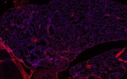
- Immunohistochemistry analysis of Lrp-1b (CD91beta) in mouse E16.5 lung sections. Antigen retrieval from 4% PFA, paraffin embedded sections was performed using EDTA. Following antigen retrieval, tissues were blocked and probed with a Lrp-1b mouse monoclonal antibody (Product # 37-7600) at a dilution of 1:1000. Detection was performed using an Alexa Fluor-568-conjugated anti-mouse secondary antibody (Product # A-21124).

Supportive validation
- Submitted by
- Invitrogen Antibodies (provider)
- Main image

- Experimental details
- NULL
- Submitted by
- Invitrogen Antibodies (provider)
- Main image

- Experimental details
- NULL
- Submitted by
- Invitrogen Antibodies (provider)
- Main image
- Experimental details
- NULL
- Submitted by
- Invitrogen Antibodies (provider)
- Main image

- Experimental details
- NULL
- Submitted by
- Invitrogen Antibodies (provider)
- Main image
- Experimental details
- NULL
- Submitted by
- Invitrogen Antibodies (provider)
- Main image
- Experimental details
- NULL

- Submitted by
- Invitrogen Antibodies (provider)
- Main image
- Experimental details
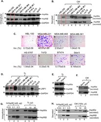
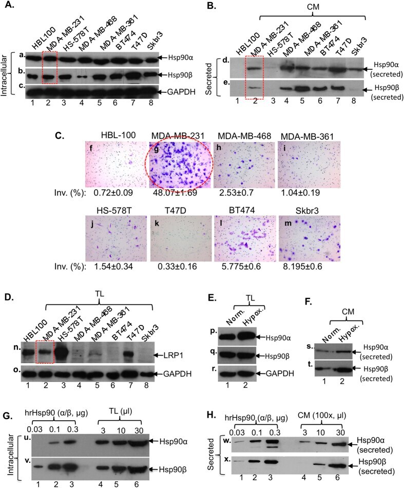
- Figure 1 Selection of MDA-MB-231 breast cancer cell line as the model of study. Western blots for Hsp90alpha and Hsp90beta in total lysates (TL) ( A ) or conditioned media (100x) (CM) ( B ) and the invasiveness ( C ) of the indicated breast cancer and control cell lines. Western blots for LRP-1 receptor among cell lines ( D ) and the total ( E ) and the secreted ( F ) Hsp90alpha and Hsp90beta under hypoxia in MDA-MB-231. Intracellular ( G ) and secreted ( H ) ratios between Hsp90alpha and Hsp90beta in MDA-MB-231 cells.
- Submitted by
- Invitrogen Antibodies (provider)
- Main image

- Experimental details
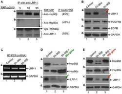
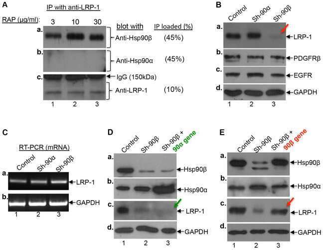
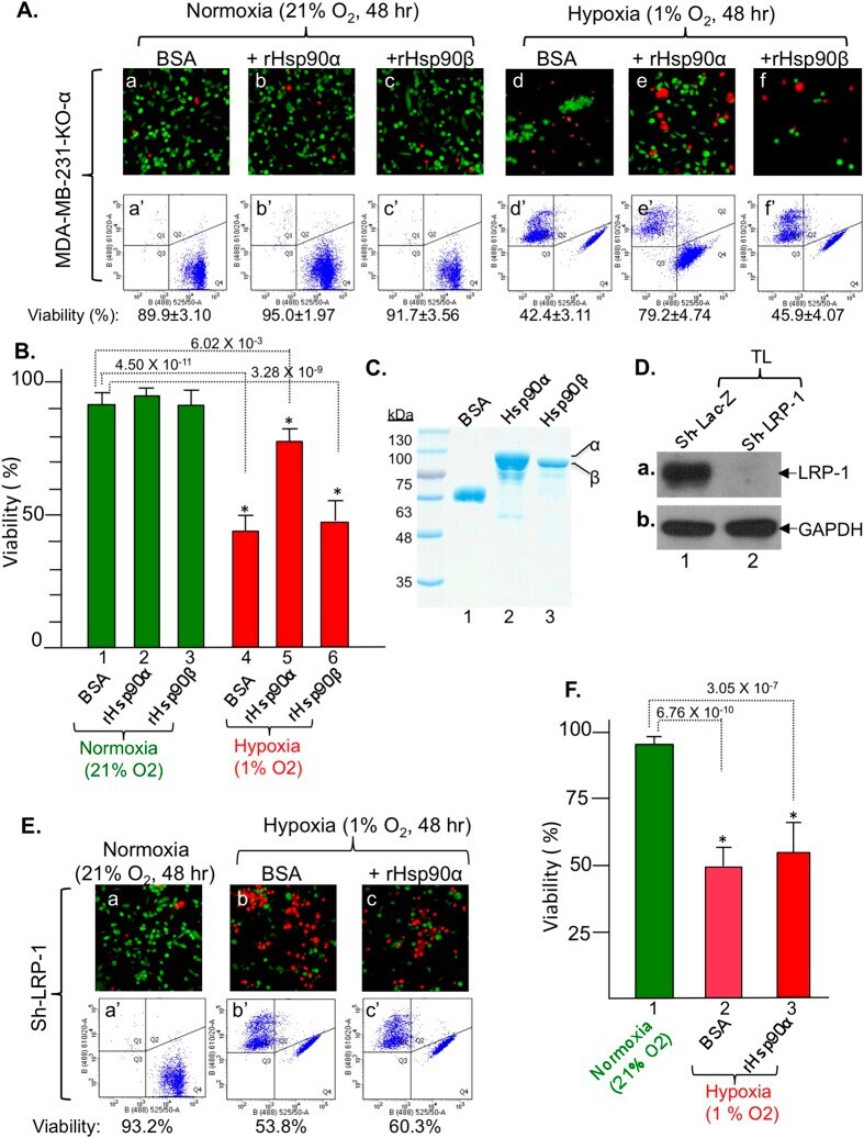
- Figure 3 Rescue of Hsp90alpha-knockout cells from hypoxia-driven killing by extracellular Hsp90alpha, but not Hsp90beta, protein via LRP-1 receptor signaling. ( A ) Extracellular Hsp90alpha, not Hsp90beta, rescues viability of Hsp90alpha-knockout cells. ( B ) Quantitation of viability data. ( C ) Evidence of purified recombinant proteins for rescues. ( D ) Down-regulation of LRP-1 shown by Western blot. ( E ) No rescue of LRP-1-downregulated cells from hypoxia by extracellular Hsp90alpha. ( F ) Quantitation . n = 3, *p < 0.05.
- Submitted by
- Invitrogen Antibodies (provider)
- Main image
- Experimental details
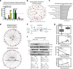
- FIGURE 7 The M7 module is enriched in activated astrocyte derived EV markers and involved in inflammatory processes. (a) The enrichment of proteins, identified as EV markers for the activated astrocytes, was calculated for each module in the AD protein network using a one-tailed Fisher's exact test. Both the raw p -value and the FDR p -value (correction for multiple comparisons by the Benjamini-Hochberg method) are shown; The dashed line represents a p -value
