 Explore
 Validate
 Learn
Learn12-0919-42
antibody from Invitrogen Antibodies
Targeting: LRP1
A2MR, APOER, APR, CD91, IGFBP-3R, IGFBP3R1, LRP, LRP1A
 Flow cytometry
Antibody data
- Antibody Data
- Antigen structure
- References [7]
- Comments [0]
- Validations
- Flow cytometry [2]
- Other assay [1]
Submit
Validation data
Reference
Comment
Report error
- Product number
- 12-0919-42 - Provider product page

- Provider
- Invitrogen Antibodies
- Product name
- CD91 Monoclonal Antibody (A2MR-a2), PE, eBioscience™
- Antibody type
- Monoclonal
- Antigen
- Other
- Description
- Description: The A2MR-a2 monoclonal antibody reacts with human CD91, also known as LRP-1 and alpha-2 macroglobulin receptor. CD91 is a 600 kDa, type 1 transmembrane protein consisting of an alpha and beta chain. The alpha chain interacts with multiple structurally and functionally diverse ligands and the beta chain contains 2 tyrosine phosphorylation motifs capable of interacting with adaptor and signaling proteins. Binding of ligands mediates ligand endocytosis and trafficking to lysosomes. This receptor is expressed by cells in the monocytic lineage, dendritic cells, hepatocytes, firbroblasts, adipocytes, neurons, and syncytiotrophoblast. CD91 is involved in cross-presenting antigens via MHC class I molecules on antigen presenting cells after endocytosis of heat shock protein-peptide complexes. It also plays a role in cell migration and is involved in activating signaling cascades. Applications Reported: This A2MR-a2 antibody has been reported for use in flow cytometric analysis. Applications Tested: This A2MR-a2 antibody has been pre-titrated and tested by flow cytometric analysis of normal human peripheral blood cells. This can be used at 5 µL (0.06 µg) per test. A test is defined as the amount (µg) of antibody that will stain a cell sample in a final volume of 100 µL. Cell number should be determined empirically but can range from 10^5 to 10^8 cells/test. Excitation: 488-561 nm; Emission: 578 nm; Laser: Blue Laser, Green Laser, Yellow-Green Laser. Filtration: 0.2 µm post-manufacturing filtered.
- Reactivity
- Human
- Host
- Mouse
- Conjugate
- Yellow dye
- Isotype
- IgG
- Antibody clone number
- A2MR-a2
- Vial size
- 100 Tests
- Concentration
- 5 µL/Test
- Storage
- 4° C, store in dark, DO NOT FREEZE!
Submitted references Accelerated Wound Healing by Fibroblasts Differentiated from Human Embryonic Stem Cell-Derived Mesenchymal Stem Cells in a Pressure Ulcer Animal Model.
An AXL/LRP-1/RANBP9 complex mediates DC efferocytosis and antigen cross-presentation in vivo.
LRP-1: a checkpoint for the extracellular matrix proteolysis.
CD91-dependent programming of T-helper cell responses following heat shock protein immunization.
Identification of the receptor scavenging hemopexin-heme complexes.
All for CD91 and CD91 for all.
CD91 is a common receptor for heat shock proteins gp96, hsp90, hsp70, and calreticulin.
Yoon D, Yoon D, Sim H, Hwang I, Lee JS, Chun W
Stem cells international 2018;2018:4789568
Stem cells international 2018;2018:4789568
An AXL/LRP-1/RANBP9 complex mediates DC efferocytosis and antigen cross-presentation in vivo.
Subramanian M, Hayes CD, Thome JJ, Thorp E, Matsushima GK, Herz J, Farber DL, Liu K, Lakshmana M, Tabas I
The Journal of clinical investigation 2014 Mar;124(3):1296-308
The Journal of clinical investigation 2014 Mar;124(3):1296-308
LRP-1: a checkpoint for the extracellular matrix proteolysis.
Etique N, Verzeaux L, Dedieu S, Emonard H
BioMed research international 2013;2013:152163
BioMed research international 2013;2013:152163
CD91-dependent programming of T-helper cell responses following heat shock protein immunization.
Pawaria S, Binder RJ
Nature communications 2011 Nov 1;2:521
Nature communications 2011 Nov 1;2:521
Identification of the receptor scavenging hemopexin-heme complexes.
Hvidberg V, Maniecki MB, Jacobsen C, Højrup P, Møller HJ, Moestrup SK
Blood 2005 Oct 1;106(7):2572-9
Blood 2005 Oct 1;106(7):2572-9
All for CD91 and CD91 for all.
Stebbing J, Savage P, Patterson S, Gazzard B
The Journal of antimicrobial chemotherapy 2004 Jan;53(1):1-3
The Journal of antimicrobial chemotherapy 2004 Jan;53(1):1-3
CD91 is a common receptor for heat shock proteins gp96, hsp90, hsp70, and calreticulin.
Basu S, Binder RJ, Ramalingam T, Srivastava PK
Immunity 2001 Mar;14(3):303-13
Immunity 2001 Mar;14(3):303-13
No comments: Submit comment
Supportive validation
- Submitted by
- Invitrogen Antibodies (provider)
- Main image
- Experimental details
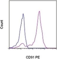
- Staining of normal human peripheral blood cells with Mouse IgG1 K Isotype Control PE (Product # 12-4714-81) (blue histogram) or Anti-Human CD91 PE (purple histogram). Cells in the monocyte gate were used for analysis.
- Conjugate
- Yellow dye
- Submitted by
- Invitrogen Antibodies (provider)
- Main image

- Experimental details
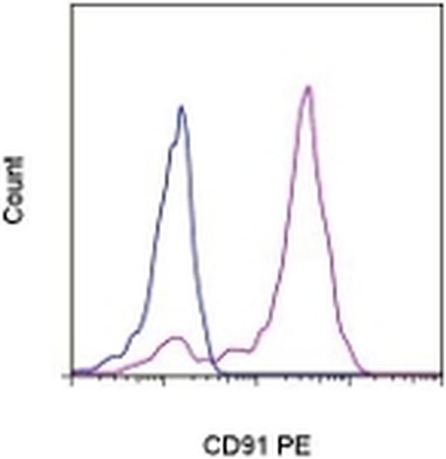
- Normal human peripheral blood cells were stained with Mouse IgG1 kappa Isotype Control, PE (Product # 12-4714-82) (blue histogram) or CD91 Monoclonal Antibody, PE (purple histogram). Cells in the monocyte gate were used for analysis.
- Conjugate
- Yellow dye

Supportive validation
- Submitted by
- Invitrogen Antibodies (provider)
- Main image
- Experimental details
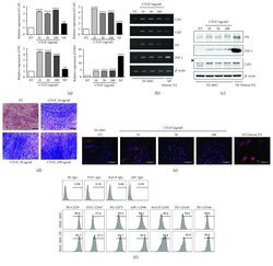
- Figure 1 Fibrogenic differentiation of human embryonic stem cell-derived mesenchymal stem cells (hESC-MSCs) upon stimulation with connective tissue growth factor (CTGF). hESC-MSCs were differentiated into fibroblasts by treatment with various concentrations of connective tissue growth factor (CTGF) for 4 weeks. Normal skin fibroblasts (Detroit 551) were also used as a positive control. (a) mRNA levels of fibroblast-related genes in hESC-MSCs after CTGF treatment were determined by the real-time polymerase chain reaction (PCR) ( n = 3, one-way ANOVA; ** p < 0.01 and **** p < 0.0001). (b) Collagen (Col)1, Col3, fibronectin (FN), and fibroblast-specific protein- (FSP-) 1 mRNA levels were determined by PCR. (c) FN, FSP-1, Col1, and beta -actin protein levels in hESC-MSCs following CTGF treatment were determined by immunoblotting. (d) Masson's trichrome was used to detect collagen fibers. (e) hESC-MSCs were immunostained to detect collagen I (Col1) following CTGF treatment. 4',6'-Diamidino-2-phenylindole (DAPI) was used for nuclear counterstaining. (f) Flow cytometry analysis of hESC-MSCs. After expansion of hESC-MSCs and hESC-MSC-Fbs, cells were trypsinized and stained with specific markers for CD29, CD47, CD73, CD90, CD91, CD105, and CD166.
- Conjugate
- Yellow dye
