 Explore
 Validate
 Learn
Learn Western blot
Antibody data
- Antibody Data
- Antigen structure
- References [0]
- Comments [0]
- Validations
- Western blot [2]
- Immunocytochemistry [1]
- Immunohistochemistry [1]
Submit
Validation data
Reference
Comment
Report error
- Product number
- 710580 - Provider product page

- Provider
- Invitrogen Antibodies
- Product name
- Troponin I Recombinant Polyclonal Antibody (1HCLC)
- Antibody type
- Polyclonal
- Antigen
- Other
- Description
- Recombinant rabbit polyclonal antibodies are unique offerings from Thermo Fisher Scientific. They are comprised of a selection of multiple different recombinant monoclonal antibodies, providing the best of both worlds - the sensitivity of polyclonal antibodies with the specificity of monoclonal antibodies - all delivered with the consistency only found in a recombinant antibody. While functionally the same as a polyclonal antibody - recognizing multiple epitope sites on the target and producing higher detection sensitivity for low abundance targets - a recombinant rabbit polyclonal antibody has a known mixture of light and heavy chains. The exact population can be produced in every lot, circumventing the biological variability typically associated with polyclonal antibody production.
- Reactivity
- Human, Mouse, Rat
- Host
- Rabbit
- Isotype
- IgG
- Antibody clone number
- 1HCLC
- Vial size
- 100 µg
- Concentration
- 0.5 mg/mL
- Storage
- Store at 4°C short term. For long term storage, store at -20°C, avoiding freeze/thaw cycles.
No comments: Submit comment
Supportive validation
- Submitted by
- Invitrogen Antibodies (provider)
- Main image
- Experimental details
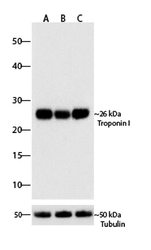
- Western blot analysis of Troponin I was performed by loading 30 µg of Rat Heart, Mouse Heart and Human Cardiomyocyte lysates (lane A, B, C) using Novex®NuPAGE®4-12% Bis-Tris gel (Product # NP0321BOX), Xcell SureLock Electrophoresis system (Product # EI0002), Novex sharp Pre-stained Protein Standard (Product # LC5800), and iBlot® Dry Blotting System (Product # IB21001). Proteins were transferred to a nitrocellulose membrane and blocked with 5% skim milk for 1 hour at room temperature. Troponin I was detected at ~26 kDa using Troponin I Recombinant Rabbit Polyclonal Antibody (Product # 710580) at a 1:1000 dilution in 2.5% skim milk at 4° C overnight on a rocking platform. Goat anti-Rabbit IgG - HRP Secondary Antibody (Product # G-21234) at 1:5000 dilution was used and chemiluminescent detection was performed using Pierce™ ECL Western blotting Substrate (Product # 32106).

- Submitted by
- Invitrogen Antibodies (provider)
- Main image

- Experimental details
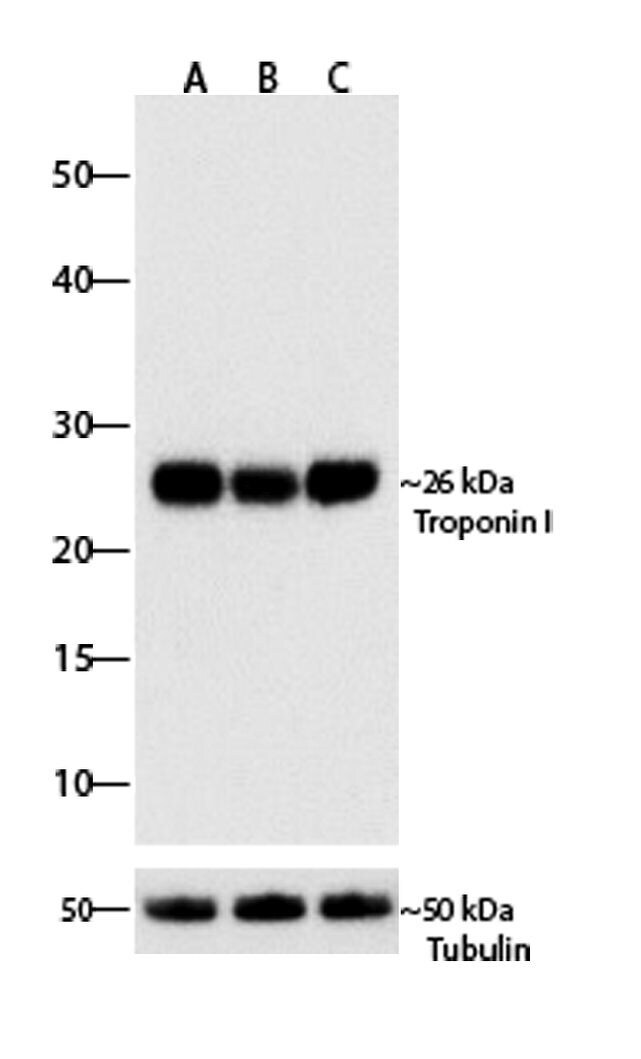
- Western blot analysis of Troponin I was performed by loading 30 µg of Rat Heart, Mouse Heart and Human Cardiomyocyte lysates (lane A, B, C) using Novex®NuPAGE®4-12% Bis-Tris gel (Product # NP0321BOX), Xcell SureLock Electrophoresis system (Product # EI0002), Novex sharp Pre-stained Protein Standard (Product # LC5800), and iBlot® Dry Blotting System (Product # IB21001). Proteins were transferred to a nitrocellulose membrane and blocked with 5% skim milk for 1 hour at room temperature. Troponin I was detected at ~26 kDa using Troponin I Recombinant Rabbit Polyclonal Antibody (Product # 710580) at a 1:1000 dilution in 2.5% skim milk at 4° C overnight on a rocking platform. Goat anti-Rabbit IgG - HRP Secondary Antibody (Product # G-21234) at 1:5000 dilution was used and chemiluminescent detection was performed using Pierce™ ECL Western blotting Substrate (Product # 32106).
Supportive validation
- Submitted by
- Invitrogen Antibodies (provider)
- Main image
- Experimental details
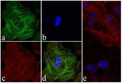
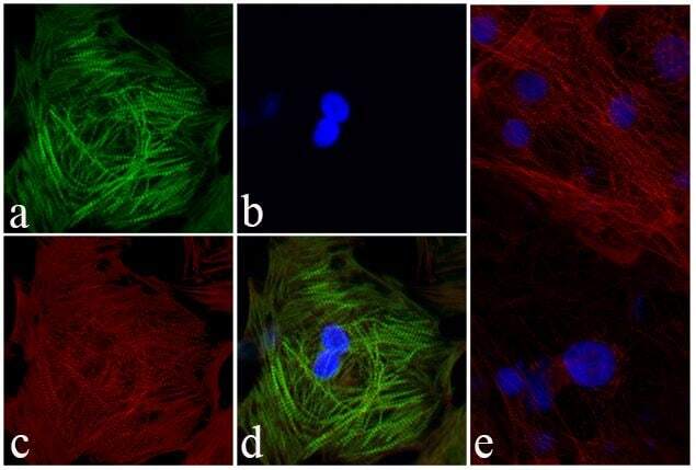
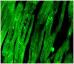
- Immunofluorescent analysis of Troponin I was done on Primary Human Cardiomyocytes. The cells were fixed with 4% paraformaldehyde for 15 minutes, permeabilized with 0.25% Triton X-100 for 10 minutes, and blocked with 5% BSA for 1 hour at room temperature. The cells were labeled with Troponin I Recombinant Rabbit Polyclonal Antibody (Product # 710580) at a dilution of 1:400 in 1% BSA and incubated for 3 hours at room temperature and then labeled with Alexa Fluor® 488 Goat anti-Rabbit IgG Secondary Antibody (Product # A-11008) at a dilution of 1:400 for 30 minutes at room temperature (Panel a: green). Nuclei (Panel b: blue) were stained with SlowFade® Gold Antifade Mountant with DAPI (Product # S36938). F-actin (Panel c: red) was stained with Alexa Fluor® 594 Phalloidin (Product # A12381). Panel d is a merged image showing striated muscle fiber localization. Panel e is no primary antibody control. The images were captured using a Nikon microscope at 20X magnification.
Supportive validation
- Submitted by
- Invitrogen Antibodies (provider)
- Main image
- Experimental details
- Immunohistochemistry analysis of Troponin I Recombinant Rabbit Polyclonal Antibody (Product # 710580) was done on paraffin embedded mouse heart tissue sections. To expose target proteins, heat induced epitope retrieval was performed using Tris-EDTA (pH 9.0) buffer for 15 minutes. Following antigen retrieval, tissues were blocked in 0.2% BSA with 0.1% cold fish skin gelatin in 1X PBS for 1 hour in a humidified chamber. The tissues were then probed at a dilution of 1:100 with Troponin I Recombinant Rabbit Polyclonal Antibody (Product # 710580) or blocking buffer alone as negative control for 3 hours at room temperature in a humidified chamber. Detection was performed using Alexa Fluor® 488 Goat anti-Rabbit IgG Secondary Antibody (Product # A-11008) at a dilution of 1:400 for 30 minutes in a humidified chamber. Tissues were counterstained with SlowFade® Gold Antifade Mountant with DAPI (Product # S36938). The images were captured using a Nikon microscope at 20X magnification.
