 Explore
 Validate
 Learn
Learn Western blot
 Immunohistochemistry
Antibody data
- Antibody Data
- Antigen structure
- References [1]
- Comments [0]
- Validations
- Immunohistochemistry [1]
Submit
Validation data
Reference
Comment
Report error
- Product number
- BAF965 - Provider product page

- Provider
- R&D Systems
- Product name
- Mouse Cathepsin B Biotinylated Antibody
- Antibody type
- Polyclonal
- Description
- Antigen Affinity-purified. Detects mouse Cathepsin B in Western blots. In Western blots, approximately 50% cross-reactivity with recombinant human Cathepsin B is observed and 15% cross-reactivity with recombinant mouse (rm) Cathepsin A, rmCathepsin C, and rmCathepsin D is observed.
- Reactivity
- Mouse
- Host
- Goat
- Conjugate
- Biotin
- Antigen sequence
P10605- Isotype
- IgG
- Vial size
- 50 ug
- Concentration
- LYOPH
- Storage
- Use a manual defrost freezer and avoid repeated freeze-thaw cycles. 12 months from date of receipt, -20 to -70 °C as supplied. 1 month, 2 to 8 °C under sterile conditions after reconstitution. 6 months, -20 to -70 °C under sterile conditions after reconstitution.
Submitted references Cathepsin L proteolytically processes histone H3 during mouse embryonic stem cell differentiation.
Duncan EM, Muratore-Schroeder TL, Cook RG, Garcia BA, Shabanowitz J, Hunt DF, Allis CD
Cell 2008 Oct 17;135(2):284-94
Cell 2008 Oct 17;135(2):284-94
No comments: Submit comment
Supportive validation
- Submitted by
- R&D Systems (provider)
- Main image
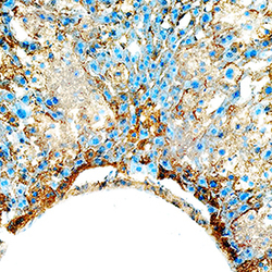
- Experimental details
- Cathepsin B in Mouse Liver. Cathepsin B was detected in perfusion fixed frozen sections of mouse liver using Goat Anti-Mouse Cathepsin B Biotinylated Antigen Affinity-purified Polyclonal Antibody (Catalog # BAF965) at 15 µg/mL overnight at 4 °C. Tissue was stained using the Anti-Goat HRP-DAB Cell & Tissue Staining Kit (brown; Catalog # CTS008) and counterstained with hematoxylin (blue). Specific staining was localized to cytoplasm in hepatocytes. View our protocol for Chromogenic IHC Staining of Frozen Tissue Sections.
