 Explore
 Validate
 Learn
Learn Western blot
Antibody data
- Antibody Data
- Antigen structure
- References [3]
- Comments [0]
- Validations
- Western blot [3]
- Immunohistochemistry [9]
- Other assay [2]
Submit
Validation data
Reference
Comment
Report error
- Product number
- MA5-32651 - Provider product page

- Provider
- Invitrogen Antibodies
- Product name
- Cathepsin B Recombinant Rabbit Monoclonal Antibody (JA11-02)
- Antibody type
- Monoclonal
- Antigen
- Recombinant full-length protein
- Description
- Recombinant rabbit monoclonal antibodies are produced using in vitro expression systems. The expression systems are developed by cloning in the specific antibody DNA sequences from immunoreactive rabbits. Then, individual clones are screened to select the best candidates for production. The advantages of using recombinant rabbit monoclonal antibodies include: better specificity and sensitivity, lot-to-lot consistency, animal origin-free formulations, and broader immunoreactivity to diverse targets due to larger rabbit immune repertoire.
- Reactivity
- Human
- Host
- Rabbit
- Isotype
- IgG
- Antibody clone number
- JA11-02
- Vial size
- 100 μL
- Concentration
- 1 mg/mL
- Storage
- Store at 4°C short term. For long term storage, store at -20°C, avoiding freeze/thaw cycles.
Submitted references SARS-CoV-2 Permissive glioblastoma cell line for high throughput antiviral screening.
ANP and BNP Exert Anti-Inflammatory Action via NPR-1/cGMP Axis by Interfering with Canonical, Non-Canonical, and Alternative Routes of Inflammasome Activation in Human THP1 Cells.
Extracellular Vesicles from Human Advanced-Stage Prostate Cancer Cells Modify the Inflammatory Response of Microenvironment-Residing Cells.
Vanhulle E, Stroobants J, Provinciael B, Camps A, Noppen S, Maes P, Vermeire K
Antiviral research 2022 Jul;203:105342
Antiviral research 2022 Jul;203:105342
ANP and BNP Exert Anti-Inflammatory Action via NPR-1/cGMP Axis by Interfering with Canonical, Non-Canonical, and Alternative Routes of Inflammasome Activation in Human THP1 Cells.
Mezzasoma L, Talesa VN, Romani R, Bellezza I
International journal of molecular sciences 2020 Dec 22;22(1)
International journal of molecular sciences 2020 Dec 22;22(1)
Extracellular Vesicles from Human Advanced-Stage Prostate Cancer Cells Modify the Inflammatory Response of Microenvironment-Residing Cells.
Mezzasoma L, Costanzi E, Scarpelli P, Talesa VN, Bellezza I
Cancers 2019 Aug 30;11(9)
Cancers 2019 Aug 30;11(9)
No comments: Submit comment
Supportive validation
- Submitted by
- Invitrogen Antibodies (provider)
- Main image
- Experimental details
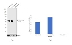
- Knockdown of Cathepsin B was achieved by transfecting MCF7 with Cathepsin B specific siRNAs (Silencer® select Product # S3739, S3738). Western blot analysis (Fig. a) was performed using Whole cell extracts from the Cathepsin B knockdown cells (lane 3), non-targeting scrambled siRNA transfected cells (lane 2) and untransfected cells (lane 1). The blot was probed with Cathepsin B Recombinant Rabbit Monoclonal Antibody (JA11-02) (Product # MA5-32651, 1:1000 dilution) and Goat anti-Rabbit IgG (Heavy Chain) Superclonal™ Recombinant Secondary Antibody, HRP (Product # A27036, 1:4000 dilution). Densitometric analysis of this western blot is shown in histogram (Fig. b). Decrease in signal upon siRNA mediated knock down confirms that antibody is specific to Pro-cathepsin B.

- Submitted by
- Invitrogen Antibodies (provider)
- Main image
- Experimental details
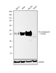
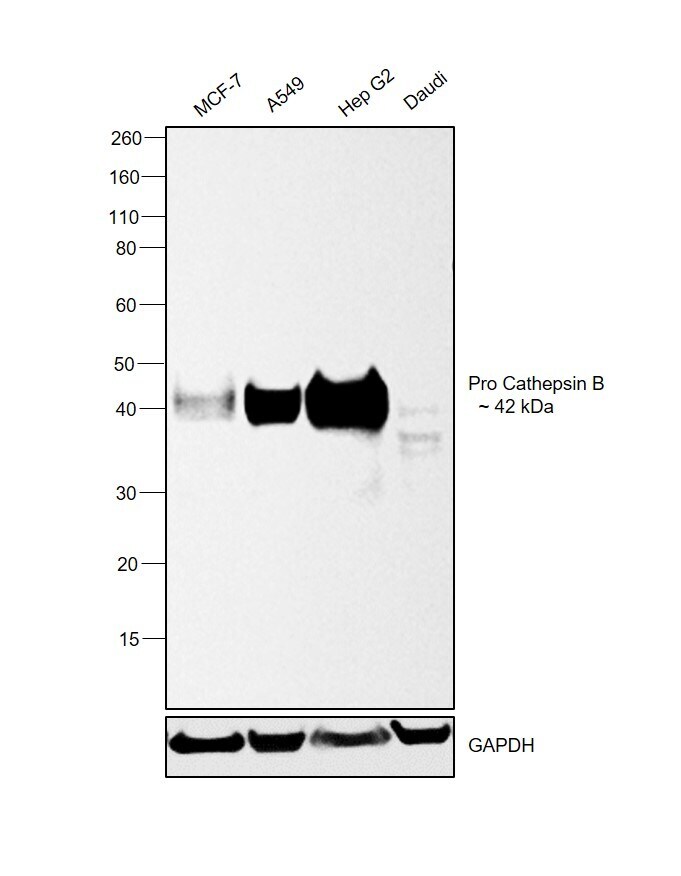
- Western blot was performed using Anti-Cathepsin B Recombinant Rabbit Monoclonal Antibody (JA11-02)(Product # MA5-32651) and a 42kDa band corresponding to Pro Cathepsin B was observed across cell lines tested except Daudi which is reported to be low. Whole cell extracts (60 µg lysate) of MCF7 (Lane 1), A549 (Lane 2), Hep G2 (Lane 3) and Daudi (Lane 4) were electrophoresed using NuPAGE™ 4-12% Bis-Tris Protein Gel (Product # NP0321BOX). Resolved proteins were then transferred onto a Nitrocellulose membrane (Product # IB23001) by iBlot® 2 Dry Blotting System (Product # IB21001). The blot was probed with the primary antibody (1:1000 dilution) and detected by chemiluminescence with Goat anti-Rabbit IgG (Heavy Chain) Superclonal™ Recombinant Secondary Antibody, HRP (Product # A27036, 1:4000 dilution) using the iBright FL 1000 (Product # A32752). Chemiluminescent detection was performed using SuperSignal™ West Dura Extended Duration Substrate (Product # 34076).
- Submitted by
- Invitrogen Antibodies (provider)
- Main image
- Experimental details
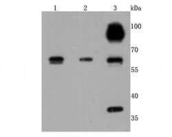
- Western blot analysis of Cathepsin B in different lysates using a Monoclonal antibody (Product #MA5-32651) at a dilution of 1:500. Positive control: Lane 1: HepG2, Lane 2: Hela, Lane 3:A549.

Supportive validation
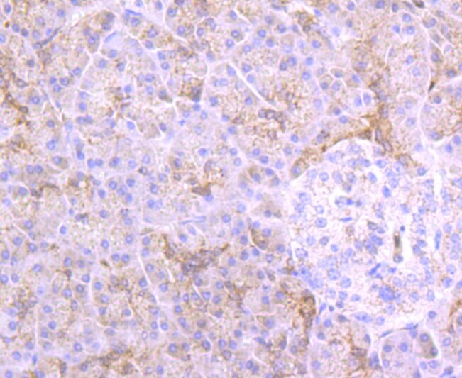
- Submitted by
- Invitrogen Antibodies (provider)
- Main image
- Experimental details
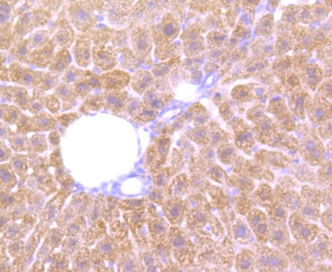
- Immunohistochemical analysis of Cathepsin B of paraffin-embedded Human liver cancer tissue using a Cathepsin-B Monoclonal antibody (Product #MA5-32651). Counter stained with hematoxylin.

- Submitted by
- Invitrogen Antibodies (provider)
- Main image
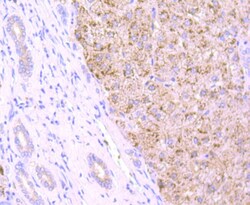
- Experimental details
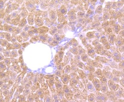
- Immunohistochemical analysis of Cathepsin B of paraffin-embedded Human liver tissue using a Cathepsin-B Monoclonal antibody (Product #MA5-32651). Counter stained with hematoxylin.

- Submitted by
- Invitrogen Antibodies (provider)
- Main image
- Experimental details
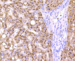
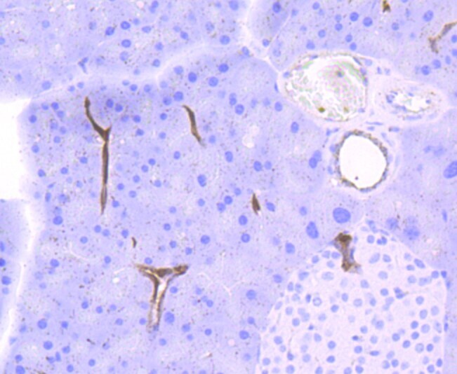
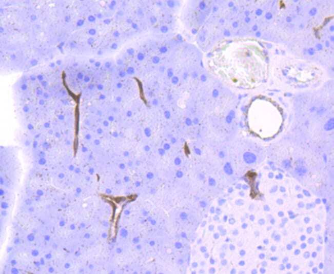
- Immunohistochemical analysis of Cathepsin B of paraffin-embedded Human pancreas tissue using a Cathepsin-B Monoclonal antibody (Product #MA5-32651). Counter stained with hematoxylin.
- Submitted by
- Invitrogen Antibodies (provider)
- Main image

- Experimental details
- Immunohistochemical analysis of Cathepsin B of paraffin-embedded Mouse liver tissue using a Cathepsin-B Monoclonal antibody (Product #MA5-32651). Counter stained with hematoxylin.
- Submitted by
- Invitrogen Antibodies (provider)
- Main image

- Experimental details
- Immunohistochemical analysis of Cathepsin B of paraffin-embedded Mouse pancreas tissue using a Cathepsin-B Monoclonal antibody (Product #MA5-32651). Counter stained with hematoxylin.
- Submitted by
- Invitrogen Antibodies (provider)
- Main image

- Experimental details
- Immunohistochemical analysis of Cathepsin B of paraffin-embedded Mouse brain tissue using a Cathepsin-B Monoclonal antibody (Product #MA5-32651). Counter stained with hematoxylin.

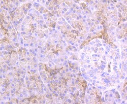
- Submitted by
- Invitrogen Antibodies (provider)
- Main image

- Experimental details
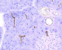
- Immunohistochemical analysis of Cathepsin B of paraffin-embedded Human pancreas tissue using a Cathepsin-B Monoclonal antibody (Product #MA5-32651). Counter stained with hematoxylin.

- Submitted by
- Invitrogen Antibodies (provider)
- Main image
- Experimental details
- Immunohistochemical analysis of Cathepsin B of paraffin-embedded Mouse liver tissue using a Cathepsin-B Monoclonal antibody (Product #MA5-32651). Counter stained with hematoxylin.

- Submitted by
- Invitrogen Antibodies (provider)
- Main image
- Experimental details
- Immunohistochemical analysis of Cathepsin B of paraffin-embedded Mouse pancreas tissue using a Cathepsin-B Monoclonal antibody (Product #MA5-32651). Counter stained with hematoxylin.
Supportive validation
- Submitted by
- Invitrogen Antibodies (provider)
- Main image
- Experimental details
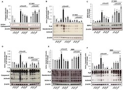
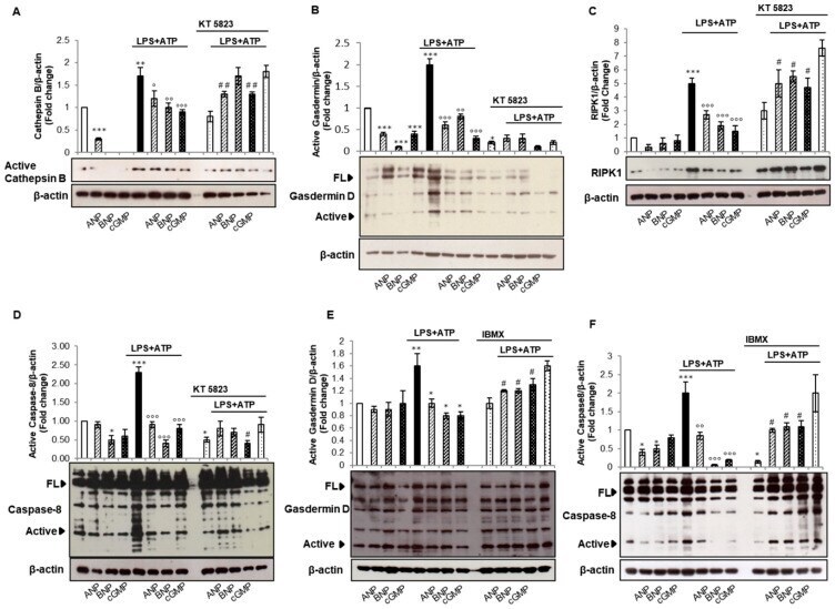
- Figure 5 cGMP but not PKG-I is involved in non-canonical and alternative inflammasome activation. THP-1 cells were pre-treated for 10 min with ANP (10 -8 M) or BNP (10 -8 M) or cGMP (100 µM 8-Br-cGMP) in the absence or presence of LPS (10 µg/mL for 20 min) + ATP (5 mM for 40 min). When KT-5823 (1 muM) or 3-isobutyl-1-methylxanthine (IBMX) (10 muM) were used, they were added 1 h before ANP, BNP, or 8-Br-cGMP treatment. Cell lysates were immunoblotted for active Cathepsin B ( A ), Gasdermin D ( B , E ), RIPK1 ( C ), or Caspase-8 ( D , F ). Protein loading was assessed by re-probing the blots with an anti-beta-actin antibody. One representative image is shown. Representative western blots images are shown. Histograms of densitometric quantification. Bars represent the ratio between respective protein and beta-actin band intensity. Untreated cells were used as control and assumed as 1. All histograms indicate mean +- SD of at least n = three independent experiments, each one tested in triplicate. * p < 0.05, ** p < 0.01, *** p < 0.001 versus untreated cells; deg p < 0.05, degdeg p < 0.01 and degdegdeg p < 0.001 versus LPS + ATP treated cells; # p < 0.05 versus KT-5823 + LPS + ATP treated cells.
- Submitted by
- Invitrogen Antibodies (provider)
- Main image
- Experimental details
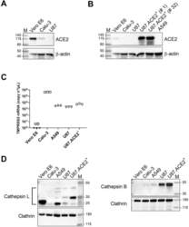
- Fig. 1 U87 cells have no detectable endogenous ACE2 expression but high levels of Cathepsin B. A) Different cell lines were analyzed for ACE2 expression by immunoblotting, with beta-actin as loading control. B) Same as in (A) but with the stably ACE2 transduced U87 cells, tested at an early (# 1) or late (# 32) passage of the cells. The transduced U87 cells stably express ACE2 at a very high level as compared to endogenous ACE2 in the Vero E6 cells. The same immunoblot is used for panel A and B, but with a longer exposure time for panel A in order to visualize the faint band in the Calu-3 sample. C) Comparative qPCR analysis of TMPRSS2 mRNA levels in different cells. Graph shows individual copy numbers/mul of 3 technical replicates (n = 3) as calculated from a TMPRSS2 standard. The TMPRSS2 level in Vero E6 cells was below detection limit. D) Different cell lines were analyzed for Cathepsin L (CTSL; left) and Cathepsin B (CTSB; right) expression by immunoblotting, with clathrin as loading control. For CTSL, different species are detected (indicated by the bracket): Pro-CTSL (41 kDa), glycosylated mature CTSL (31 kDa) and non-glycosylated mature CTSL (25 kDa), whereas only the Pro-CTSB form (42 kDa) is visualized. M: molecular marker in kDa, UD: undetectable. Fig. 1
