Explore
 Validate
 Learn
Learn Western blot
Antibody data
- Antibody Data
- Antigen structure
- References [0]
- Comments [0]
- Validations
- Western blot [4]
Submit
Validation data
Reference
Comment
Report error
- Product number
- PA5-17006 - Provider product page

- Provider
- Invitrogen Antibodies
- Product name
- Cathepsin B Polyclonal Antibody
- Antibody type
- Polyclonal
- Antigen
- Synthetic peptide
- Description
- It is not recommended to aliquot this antibody.
- Reactivity
- Human
- Host
- Rabbit
- Isotype
- IgG
- Vial size
- 100 μL
- Concentration
- 26 μg/mL
- Storage
- -20°C
No comments: Submit comment
Supportive validation
- Submitted by
- Invitrogen Antibodies (provider)
- Main image
- Experimental details
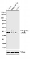
- Western blot analysis was performed on whole cell extracts (30 µg lysate) of T-47D (Lane 1), MDA-MB-231 (Lane 2) and THP-1 (Lane 3). The blot was probed with anti-Cathepsin B Rabbit Polyclonal Antibody (Product # PA5-17006, 1:500 dilution) and detected by chemiluminescence using Goat anti Rabbit IgG (Heavy Chain) Superclonal™ Secondary Antibody, HRP conjugate (Product # A27036, 0.25 µg/mL, 1:4000 dilution). A 37 kDa band corresponding to Cathepsin B was detected across the cell lines tested.

- Submitted by
- Invitrogen Antibodies (provider)
- Main image
- Experimental details
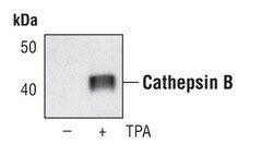
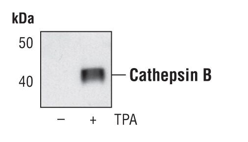
- Western blot analysis of Cathepsin B in extracts from secreted protein from HL-60 cells, untreated or treated overnight with TPA (20 nM), using Cathepsin B polyclonal antibody (Product # PA5-17006).
- Submitted by
- Invitrogen Antibodies (provider)
- Main image

- Experimental details
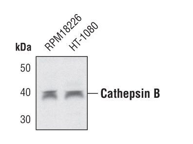
- Western blot analysis of Cathepsin B in extracts from RPM18226 and HT-1080 cell lines using Cathepsin B polyclonal antibody (Product # PA5-17006).
- Submitted by
- Invitrogen Antibodies (provider)
- Main image
- Experimental details
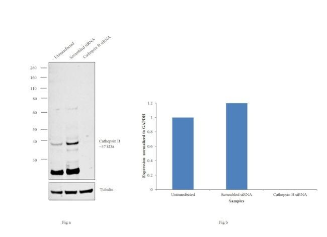
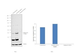
- Knockdown of Cathepsin B was achieved by transfecting MCF7 cells with Cathepsin B specific siRNAs (Silencer® select Product # s3739). Western blot analysis (Fig. a) was performed using whole cell extracts from Cathepsin B knockdown cells (lane 3), non-specific scrambled siRNA transfected cells (lane 2) and untransfected cells (lane 1). The blots were probed with Cathepsin B Polyclonal Antibody (Product # PA5-17006, 1:500 dilution) and Goat anti-Rabbit IgG (Heavy Chain) Superclonal™ Secondary Antibody, HRP conjugate (Product # A27036, 0.25 µg/mL, 1:4000 dilution). Densitometric analysis of this western blot is shown in histogram (Fig. b). Loss of signal upon siRNA mediated knock down confirms that antibody is specific to Cathepsin B.