Explore
 Validate
 Learn
Learn Western blot
 Immunoprecipitation
Antibody data
- Antibody Data
- Antigen structure
- References [15]
- Comments [0]
- Validations
- Western blot [3]
- Immunohistochemistry [1]
Submit
Validation data
Reference
Comment
Report error
- Product number
- AF953 - Provider product page

- Provider
- R&D Systems
- Product name
- Human Cathepsin B Antibody
- Antibody type
- Polyclonal
- Description
- Antigen Affinity-purified. Detects human Cathepsin B in direct ELISAs and Western blots. In direct ELISAs, approximately 35% cross-reactivity with recombinant mouse (rm) Cathepsin B is observed and less than 5% cross-reactivity with recombinant human (rh) Cathepsin C, rmCathepsin H, and rhCathepsin L is observed.
- Reactivity
- Human
- Host
- Goat
- Conjugate
- Unconjugated
- Antigen sequence
P07858- Isotype
- IgG
- Vial size
- 100 ug
- Concentration
- LYOPH
- Storage
- Use a manual defrost freezer and avoid repeated freeze-thaw cycles. 12 months from date of receipt, -20 to -70 °C as supplied. 1 month, 2 to 8 °C under sterile conditions after reconstitution. 6 months, -20 to -70 °C under sterile conditions after reconstitution.
Submitted references YWHA/14-3-3 proteins recognize phosphorylated TFEB by a noncanonical mode for controlling TFEB cytoplasmic localization.
Modulation of Receptor Protein Tyrosine Phosphatase Sigma Increases Chondroitin Sulfate Proteoglycan Degradation through Cathepsin B Secretion to Enhance Axon Outgrowth.
The lysosomal protein cathepsin L is a progranulin protease.
The Unusual Resistance of Avian Defensin AvBD7 to Proteolytic Enzymes Preserves Its Antibacterial Activity.
Cathepsin S attenuates endosomal EGFR signalling: A mechanical rationale for the combination of cathepsin S and EGFR tyrosine kinase inhibitors.
Simvastatin inhibits glucose metabolism and legumain activity in human myotubes.
Human bone marrow-derived mesenchymal stem cells suppress human glioma growth through inhibition of angiogenesis.
A possible contribution of altered cathepsin B expression to the development of skin sclerosis and vasculopathy in systemic sclerosis.
Differential secretome analysis reveals CST6 as a suppressor of breast cancer bone metastasis.
CD40 stimulation sensitizes CLL cells to lysosomal cell death induction by type II anti-CD20 mAb GA101.
Transgenic expression of human cathepsin B promotes progression and metastasis of polyoma-middle-T-induced breast cancer in mice.
Macrophages and cathepsin proteases blunt chemotherapeutic response in breast cancer.
The NALP3 inflammasome is involved in the innate immune response to amyloid-beta.
Distinct roles for cysteine cathepsin genes in multistage tumorigenesis.
Biomarker discovery from pancreatic cancer secretome using a differential proteomic approach.
Xu Y, Ren J, He X, Chen H, Wei T, Feng W
Autophagy 2019 Jun;15(6):1017-1030
Autophagy 2019 Jun;15(6):1017-1030
Modulation of Receptor Protein Tyrosine Phosphatase Sigma Increases Chondroitin Sulfate Proteoglycan Degradation through Cathepsin B Secretion to Enhance Axon Outgrowth.
Tran AP, Sundar S, Yu M, Lang BT, Silver J
The Journal of neuroscience : the official journal of the Society for Neuroscience 2018 Jun 6;38(23):5399-5414
The Journal of neuroscience : the official journal of the Society for Neuroscience 2018 Jun 6;38(23):5399-5414
The lysosomal protein cathepsin L is a progranulin protease.
Lee CW, Stankowski JN, Chew J, Cook CN, Lam YW, Almeida S, Carlomagno Y, Lau KF, Prudencio M, Gao FB, Bogyo M, Dickson DW, Petrucelli L
Molecular neurodegeneration 2017 Jul 25;12(1):55
Molecular neurodegeneration 2017 Jul 25;12(1):55
The Unusual Resistance of Avian Defensin AvBD7 to Proteolytic Enzymes Preserves Its Antibacterial Activity.
Bailleul G, Kravtzoff A, Joulin-Giet A, Lecaille F, Labas V, Meudal H, Loth K, Teixeira-Gomes AP, Gilbert FB, Coquet L, Jouenne T, Brömme D, Schouler C, Landon C, Lalmanach G, Lalmanach AC
PloS one 2016;11(8):e0161573
PloS one 2016;11(8):e0161573
Cathepsin S attenuates endosomal EGFR signalling: A mechanical rationale for the combination of cathepsin S and EGFR tyrosine kinase inhibitors.
Huang CC, Lee CC, Lin HH, Chang JY
Scientific reports 2016 Jul 8;6:29256
Scientific reports 2016 Jul 8;6:29256
Simvastatin inhibits glucose metabolism and legumain activity in human myotubes.
Smith R, Solberg R, Jacobsen LL, Voreland AL, Rustan AC, Thoresen GH, Johansen HT
PloS one 2014;9(1):e85721
PloS one 2014;9(1):e85721
Human bone marrow-derived mesenchymal stem cells suppress human glioma growth through inhibition of angiogenesis.
Ho IA, Toh HC, Ng WH, Teo YL, Guo CM, Hui KM, Lam PY
Stem cells (Dayton, Ohio) 2013 Jan;31(1):146-55
Stem cells (Dayton, Ohio) 2013 Jan;31(1):146-55
A possible contribution of altered cathepsin B expression to the development of skin sclerosis and vasculopathy in systemic sclerosis.
Noda S, Asano Y, Akamata K, Aozasa N, Taniguchi T, Takahashi T, Ichimura Y, Toyama T, Sumida H, Yanaba K, Tada Y, Sugaya M, Kadono T, Sato S
PloS one 2012;7(2):e32272
PloS one 2012;7(2):e32272
Differential secretome analysis reveals CST6 as a suppressor of breast cancer bone metastasis.
Jin L, Zhang Y, Li H, Yao L, Fu D, Yao X, Xu LX, Hu X, Hu G
Cell research 2012 Sep;22(9):1356-73
Cell research 2012 Sep;22(9):1356-73
CD40 stimulation sensitizes CLL cells to lysosomal cell death induction by type II anti-CD20 mAb GA101.
Jak M, van Bochove GG, Reits EA, Kallemeijn WW, Tromp JM, Umana P, Klein C, van Lier RA, van Oers MH, Eldering E
Blood 2011 Nov 10;118(19):5178-88
Blood 2011 Nov 10;118(19):5178-88
Transgenic expression of human cathepsin B promotes progression and metastasis of polyoma-middle-T-induced breast cancer in mice.
Sevenich L, Werner F, Gajda M, Schurigt U, Sieber C, Müller S, Follo M, Peters C, Reinheckel T
Oncogene 2011 Jan 6;30(1):54-64
Oncogene 2011 Jan 6;30(1):54-64
Macrophages and cathepsin proteases blunt chemotherapeutic response in breast cancer.
Shree T, Olson OC, Elie BT, Kester JC, Garfall AL, Simpson K, Bell-McGuinn KM, Zabor EC, Brogi E, Joyce JA
Genes & development 2011 Dec 1;25(23):2465-79
Genes & development 2011 Dec 1;25(23):2465-79
The NALP3 inflammasome is involved in the innate immune response to amyloid-beta.
Halle A, Hornung V, Petzold GC, Stewart CR, Monks BG, Reinheckel T, Fitzgerald KA, Latz E, Moore KJ, Golenbock DT
Nature immunology 2008 Aug;9(8):857-65
Nature immunology 2008 Aug;9(8):857-65
Distinct roles for cysteine cathepsin genes in multistage tumorigenesis.
Gocheva V, Zeng W, Ke D, Klimstra D, Reinheckel T, Peters C, Hanahan D, Joyce JA
Genes & development 2006 Mar 1;20(5):543-56
Genes & development 2006 Mar 1;20(5):543-56
Biomarker discovery from pancreatic cancer secretome using a differential proteomic approach.
Grønborg M, Kristiansen TZ, Iwahori A, Chang R, Reddy R, Sato N, Molina H, Jensen ON, Hruban RH, Goggins MG, Maitra A, Pandey A
Molecular & cellular proteomics : MCP 2006 Jan;5(1):157-71
Molecular & cellular proteomics : MCP 2006 Jan;5(1):157-71
No comments: Submit comment
Supportive validation
- Submitted by
- R&D Systems (provider)
- Main image
- Experimental details
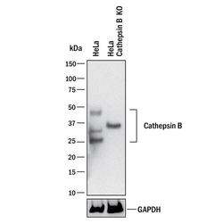
- Western Blot Shows Human Cathepsin B Specificity by Using Knockout Cell Line. Western blot shows lysates of HeLa human cervical epithelial carcinoma parental cell line and Cathepsin B knockout HeLa cell line (KO). PVDF membrane was probed with 0.25 µg/mL of Goat Anti-Human Cathepsin B Antigen Affinity-purified Polyclonal Antibody (Catalog # AF953) followed by HRP-conjugated Anti-Goat IgG Secondary Antibody (Catalog # HAF017). Specific bands were detected for Cathepsin B at approximately 25-45 kDa (as indicated) in the parental HeLa cell line, but is not detectable in knockout HeLa cell line. GAPDH (Catalog # AF5718) is shown as a loading control. This experiment was conducted under reducing conditions and using Immunoblot Buffer Group 1. New adjunct appears with knockout cell line.

- Submitted by
- R&D Systems (provider)
- Main image

- Experimental details
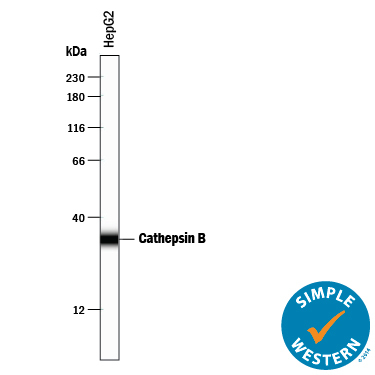
- Detection of Human Cathepsin B by Simple Western<SUP abp="263">TM. Simple Western lane view shows lysates of HepG2 human hepatocellular carcinoma cell line, loaded at 0.2 mg/mL. A specific band was detected for Cathepsin B at approximately 34 kDa (as indicated) using 2.5 µg/mL of Goat Anti-Human Cathepsin B Antigen Affinity-purified Polyclonal Antibody (Catalog # AF953) followed by 1:50 dilution of HRP-conjugated Anti-Goat IgG Secondary Antibody (Catalog # HAF109). This experiment was conducted under reducing conditions and using the 12-230 kDa separation system.
- Submitted by
- R&D Systems (provider)
- Main image

- Experimental details
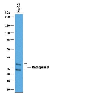
- Detection of Human Cathepsin B by Western Blot. Western blot shows lysates of HepG2 human hepatocellular carcinoma cell line. PVDF membrane was probed with 0.25 µg/mL of Goat Anti-Human Cathepsin B Antigen Affinity-purified Polyclonal Antibody (Catalog # AF953) followed by HRP-conjugated Anti-Goat IgG Secondary Antibody (Catalog # HAF019). A specific band was detected for Cathepsin B at approximately 25-30 kDa (as indicated). This experiment was conducted under reducing conditions and using Immunoblot Buffer Group 1.
Supportive validation
- Submitted by
- R&D Systems (provider)
- Main image
- Experimental details
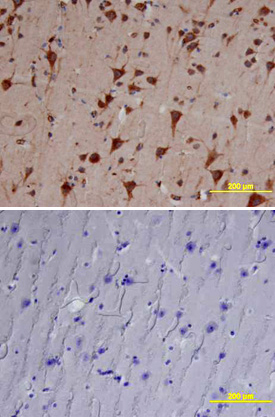
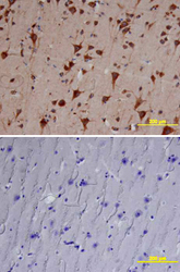
- Cathepsin B in Human Brain. Cathepsin B was detected in immersion fixed paraffin-embedded sections of human brain (cortex) using Goat Anti-Human Cathepsin B Antigen Affinity-purified Polyclonal Antibody (Catalog # AF953) at 10 µg/mL overnight at 4 °C. Tissue was stained using the Anti-Goat HRP-DAB Cell & Tissue Staining Kit (brown; Catalog # CTS008) and counterstained with hematoxylin (blue). Lower panel shows a lack of labeling if primary antibodies are omitted and tissue is stained only with secondary antibody followed by incubation with detection reagents. View our protocol for Chromogenic IHC Staining of Paraffin-embedded Tissue Sections.