Explore
 Validate
 Learn
Learn Immunocytochemistry
Antibody data
- Antibody Data
- Antigen structure
- References [5]
- Comments [0]
- Validations
- Immunocytochemistry [1]
- Immunohistochemistry [1]
Submit
Validation data
Reference
Comment
Report error
- Product number
- HPA018156 - Provider product page

- Provider
- Atlas Antibodies
- Proper citation
- Atlas Antibodies Cat#HPA018156, RRID:AB_1846069
- Product name
- Anti-CTSB
- Antibody type
- Polyclonal
- Description
- Polyclonal Antibody against Human CTSB, Gene description: cathepsin B, Validated applications: ICC, IHC, Uniprot ID: P07858, Storage: Store at +4°C for short term storage. Long time storage is recommended at -20°C.
- Reactivity
- Human
- Host
- Rabbit
- Conjugate
- Unconjugated
- Isotype
- IgG
- Vial size
- 100 µl
- Concentration
- 0.1 mg/ml
- Storage
- Store at +4°C for short term storage. Long time storage is recommended at -20°C.
- Handling
- The antibody solution should be gently mixed before use.
Submitted references Sugar transporter Slc37a2 regulates bone metabolism in mice via a tubular lysosomal network in osteoclasts.
Cathepsin B Is Upregulated and Mediates ECM Degradation in Colon Adenocarcinoma HT29 Cells Overexpressing Snail
A High-throughput Bead-based Affinity Assay Enables Analysis of Genital Protein Signatures in Women At Risk of HIV Infection
Profiling of Atherosclerotic Lesions by Gene and Tissue Microarrays Reveals PCSK6 as a Novel Protease in Unstable Carotid Atherosclerosis
Heterogeneity in signaling pathways of gastroenteropancreatic neuroendocrine tumors: a critical look at notch signaling pathway
Ng PY, Ribet ABP, Guo Q, Mullin BH, Tan JWY, Landao-Bassonga E, Stephens S, Chen K, Yuan J, Abudulai L, Bollen M, Nguyen ETTT, Kular J, Papadimitriou JM, Søe K, Teasdale RD, Xu J, Parton RG, Takayanagi H, Pavlos NJ
Nature communications 2023 Feb 21;14(1):906
Nature communications 2023 Feb 21;14(1):906
Cathepsin B Is Upregulated and Mediates ECM Degradation in Colon Adenocarcinoma HT29 Cells Overexpressing Snail
Kryczka J, Papiewska-Pajak I, Kowalska M, Boncela J
Cells 2019;8(3):203
Cells 2019;8(3):203
A High-throughput Bead-based Affinity Assay Enables Analysis of Genital Protein Signatures in Women At Risk of HIV Infection
Månberg A, Bradley F, Qundos U, Guthrie B, Birse K, Noël-Romas L, Lindskog C, Bosire R, Kiarie J, Farquhar C, Burgener A, Nilsson P, Broliden K
Molecular & Cellular Proteomics 2019;18(3):461-476
Molecular & Cellular Proteomics 2019;18(3):461-476
Profiling of Atherosclerotic Lesions by Gene and Tissue Microarrays Reveals PCSK6 as a Novel Protease in Unstable Carotid Atherosclerosis
Perisic L, Hedin E, Razuvaev A, Lengquist M, Osterholm C, Folkersen L, Gillgren P, Paulsson-Berne G, Ponten F, Odeberg J, Hedin U
Arteriosclerosis, Thrombosis, and Vascular Biology 2013;33(10):2432-2443
Arteriosclerosis, Thrombosis, and Vascular Biology 2013;33(10):2432-2443
Heterogeneity in signaling pathways of gastroenteropancreatic neuroendocrine tumors: a critical look at notch signaling pathway
Wang H, Chen Y, Fernandez-Del Castillo C, Yilmaz O, Deshpande V
Modern Pathology 2013;26(1):139-147
Modern Pathology 2013;26(1):139-147
No comments: Submit comment
Supportive validation
- Submitted by
- Atlas Antibodies (provider)
- Main image
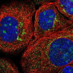
- Experimental details
- Immunofluorescent staining of human cell line A-431 shows localization to nucleoli & vesicles.
- Sample type
- Human

Supportive validation
- Submitted by
- Atlas Antibodies (provider)
- Enhanced method
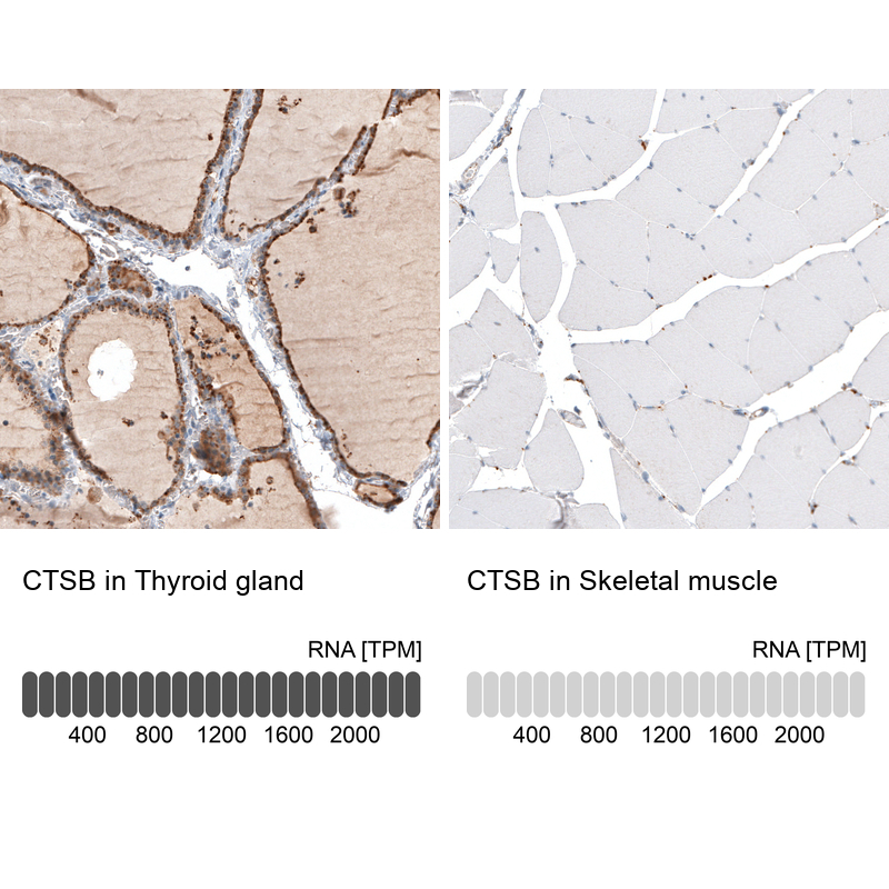
- Orthogonal validation
- Main image
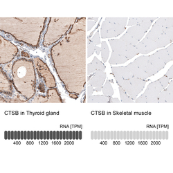
- Experimental details
- Immunohistochemistry analysis in human thyroid gland and skeletal muscle tissues using HPA018156 antibody. Corresponding CTSB RNA-seq data are presented for the same tissues.
- Sample type
- Human
- Protocol
- Protocol