 Explore
 Validate
 Learn
Learn Western blot
Antibody data
- Antibody Data
- Antigen structure
- References [14]
- Comments [0]
- Validations
- Western blot [2]
- Immunohistochemistry [2]
Submit
Validation data
Reference
Comment
Report error
- Product number
- AF1515 - Provider product page

- Provider
- Novus Biologicals
- Proper citation
- Novus Cat#AF1515, RRID:AB_2665930
- Product name
- Goat Polyclonal Cathepsin L Antibody
- Antibody type
- Polyclonal
- Description
- Antigen Affinity-purified. Detects mouse Cathepsin L in direct ELISAs and Western blots. In direct ELISAs, approximately 25% cross-reactivity with recombinant human Cathepsin L is observed.
- Reactivity
- Mouse, Rat
- Host
- Goat
- Conjugate
- Unconjugated
- Isotype
- IgG
- Vial size
- 100 ug
- Concentration
- LYOPH
- Storage
- Use a manual defrost freezer and avoid repeated freeze-thaw cycles. 12 months from date of receipt, -20 to -70 degreesC as supplied. 1 month, 2 to 8 degreesC under sterile conditions after reconstitution. 6 months, -20 to -70 degreesC under sterile conditions after reconstitution.
Submitted references Sequential, but not Concurrent, Incubation of Cathepsin K and L with Type I Collagen Results in Extended Proteolysis.
Early lysosomal maturation deficits in microglia triggers enhanced lysosomal activity in other brain cells of progranulin knockout mice.
Stat3-mediated alterations in lysosomal membrane protein composition.
Direct Observation of Enhanced Nitric Oxide in a Murine Model of Diabetic Nephropathy.
Investigating the Life Expectancy and Proteolytic Degradation of Engineered Skeletal Muscle Biological Machines.
The Role of Heparanase in the Pathogenesis of Acute Pancreatitis: A Potential Therapeutic Target.
Deficiency for the cysteine protease cathepsin L impairs Myc-induced tumorigenesis in a mouse model of pancreatic neuroendocrine cancer.
Vacuolar ATPase in phagosome-lysosome fusion.
Lysosomal protein turnover contributes to the acquisition of TGFβ-1 induced invasive properties of mammary cancer cells.
The PI3K regulatory subunits p55α and p50α regulate cell death in vivo.
Biogenesis and proteolytic processing of lysosomal DNase II.
Macrophages and cathepsin proteases blunt chemotherapeutic response in breast cancer.
The impact of microRNAs on protein output.
Major role of cathepsin L for producing the peptide hormones ACTH, beta-endorphin, and alpha-MSH, illustrated by protease gene knockout and expression.
Parks AN, Nahata J, Edouard NE, Temenoff JS, Platt MO
Scientific reports 2019 Apr 1;9(1):5399
Scientific reports 2019 Apr 1;9(1):5399
Early lysosomal maturation deficits in microglia triggers enhanced lysosomal activity in other brain cells of progranulin knockout mice.
Götzl JK, Colombo AV, Fellerer K, Reifschneider A, Werner G, Tahirovic S, Haass C, Capell A
Molecular neurodegeneration 2018 Sep 4;13(1):48
Molecular neurodegeneration 2018 Sep 4;13(1):48
Stat3-mediated alterations in lysosomal membrane protein composition.
Lloyd-Lewis B, Krueger CC, Sargeant TJ, D'Angelo ME, Deery MJ, Feret R, Howard JA, Lilley KS, Watson CJ
The Journal of biological chemistry 2018 Mar 23;293(12):4244-4261
The Journal of biological chemistry 2018 Mar 23;293(12):4244-4261
Direct Observation of Enhanced Nitric Oxide in a Murine Model of Diabetic Nephropathy.
Boels MG, van Faassen EE, Avramut MC, van der Vlag J, van den Berg BM, Rabelink TJ
PloS one 2017;12(1):e0170065
PloS one 2017;12(1):e0170065
Investigating the Life Expectancy and Proteolytic Degradation of Engineered Skeletal Muscle Biological Machines.
Cvetkovic C, Ferrall-Fairbanks MC, Ko E, Grant L, Kong H, Platt MO, Bashir R
Scientific reports 2017 Jun 19;7(1):3775
Scientific reports 2017 Jun 19;7(1):3775
The Role of Heparanase in the Pathogenesis of Acute Pancreatitis: A Potential Therapeutic Target.
Khamaysi I, Singh P, Nasser S, Awad H, Chowers Y, Sabo E, Hammond E, Gralnek I, Minkov I, Noseda A, Ilan N, Vlodavsky I, Abassi Z
Scientific reports 2017 Apr 6;7(1):715
Scientific reports 2017 Apr 6;7(1):715
Deficiency for the cysteine protease cathepsin L impairs Myc-induced tumorigenesis in a mouse model of pancreatic neuroendocrine cancer.
Brindle NR, Joyce JA, Rostker F, Lawlor ER, Swigart-Brown L, Evan G, Hanahan D, Shchors K
PloS one 2015;10(4):e0120348
PloS one 2015;10(4):e0120348
Vacuolar ATPase in phagosome-lysosome fusion.
Kissing S, Hermsen C, Repnik U, Nesset CK, von Bargen K, Griffiths G, Ichihara A, Lee BS, Schwake M, De Brabander J, Haas A, Saftig P
The Journal of biological chemistry 2015 May 29;290(22):14166-80
The Journal of biological chemistry 2015 May 29;290(22):14166-80
Lysosomal protein turnover contributes to the acquisition of TGFβ-1 induced invasive properties of mammary cancer cells.
Kern U, Wischnewski V, Biniossek ML, Schilling O, Reinheckel T
Molecular cancer 2015 Feb 15;14:39
Molecular cancer 2015 Feb 15;14:39
The PI3K regulatory subunits p55α and p50α regulate cell death in vivo.
Pensa S, Neoh K, Resemann HK, Kreuzaler PA, Abell K, Clarke NJ, Reinheckel T, Kahn CR, Watson CJ
Cell death and differentiation 2014 Sep;21(9):1442-50
Cell death and differentiation 2014 Sep;21(9):1442-50
Biogenesis and proteolytic processing of lysosomal DNase II.
Ohkouchi S, Shibata M, Sasaki M, Koike M, Safig P, Peters C, Nagata S, Uchiyama Y
PloS one 2013;8(3):e59148
PloS one 2013;8(3):e59148
Macrophages and cathepsin proteases blunt chemotherapeutic response in breast cancer.
Shree T, Olson OC, Elie BT, Kester JC, Garfall AL, Simpson K, Bell-McGuinn KM, Zabor EC, Brogi E, Joyce JA
Genes & development 2011 Dec 1;25(23):2465-79
Genes & development 2011 Dec 1;25(23):2465-79
The impact of microRNAs on protein output.
Baek D, Villén J, Shin C, Camargo FD, Gygi SP, Bartel DP
Nature 2008 Sep 4;455(7209):64-71
Nature 2008 Sep 4;455(7209):64-71
Major role of cathepsin L for producing the peptide hormones ACTH, beta-endorphin, and alpha-MSH, illustrated by protease gene knockout and expression.
Funkelstein L, Toneff T, Mosier C, Hwang SR, Beuschlein F, Lichtenauer UD, Reinheckel T, Peters C, Hook V
The Journal of biological chemistry 2008 Dec 19;283(51):35652-9
The Journal of biological chemistry 2008 Dec 19;283(51):35652-9
No comments: Submit comment
Supportive validation
- Submitted by
- Novus Biologicals (provider)
- Main image

- Experimental details
- Detection of Mouse Cathepsin L by Simple WesternTM. Simple Western lane view shows lysates of mouse liver tissue and HepG2 human hepatocellular carcinoma cell line, loaded at 0.2 mg/mL. Specific bands were detected for Cathepsin L at approximately 51 and 34 kDa (as indicated) using 10 µg/mL of Goat Anti-Mouse Cathepsin L Antigen Affinity-purified Polyclonal Antibody (Catalog # AF1515) followed by 1:50 dilution of HRP-conjugated Anti-Goat IgG Secondary Antibody (Catalog # HAF109). This experiment was conducted under reducing conditions and using the 12-230 kDa separation system.

- Submitted by
- Novus Biologicals (provider)
- Main image

- Experimental details
- Detection of Mouse and Rat Cathepsin L by Western Blot. Western blot shows lysates of rat liver tissue, mouse liver tissue (wild type), and mouse liver tissue (knock out). PVDF membrane was probed with 1 µg/mL of Goat Anti-Mouse Cathepsin L Antigen Affinity-purified Polyclonal Antibody (Catalog # AF1515) followed by HRP-conjugated Anti-Goat IgG Secondary Antibody (Catalog # HAF017). Specific bands were detected for Cathepsin L at approximately 22-38 kDa (as indicated). This experiment was conducted under reducing conditions and using Immunoblot Buffer Group 1.

Supportive validation
- Submitted by
- Novus Biologicals (provider)
- Main image

- Experimental details
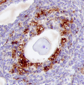
- Cathepsin L in Mouse Ovary. Cathepsin L was detected in perfusion fixed frozen sections of mouse ovary using 15 µg/mL Goat Anti-Mouse Cathepsin L Antigen Affinity-purified Polyclonal Antibody (Catalog # AF1515) overnight at 4 °C. Tissue was stained with the Anti-Goat HRP-DAB Cell & Tissue Staining Kit (brown; Catalog # CTS008) and counterstained with hematoxylin (blue). View our protocol for Chromogenic IHC Staining of Frozen Tissue Sections.
- Submitted by
- Novus Biologicals (provider)
- Main image

- Experimental details
- Cathepsin L in Mouse Thymus. Cathepsin L was detected in perfusion fixed frozen sections of mouse thymus using 15 µg/mL Goat Anti-Mouse Cathepsin L Antigen Affinity-purified Polyclonal Antibody (Catalog # AF1515) overnight at 4 °C. Tissue was stained with the Anti-Goat HRP-DAB Cell & Tissue Staining Kit (brown; Catalog # CTS0028) and counterstained with hematoxylin (blue). View our protocol for Chromogenic IHC Staining of Frozen Tissue Sections.
