Explore
 Validate
 Learn
Learn Western blot
 Immunoprecipitation
Antibody data
- Antibody Data
- Antigen structure
- References [0]
- Comments [0]
- Validations
- Western blot [1]
- Immunohistochemistry [2]
Submit
Validation data
Reference
Comment
Report error
- Product number
- MAB9521-100 - Provider product page

- Provider
- Novus Biologicals
- Product name
- Rat Monoclonal Cathepsin L Antibody
- Antibody type
- Monoclonal
- Description
- Protein A or G purified from hybridoma culture supernatant. Detects human and mouse Cathepsin L in direct ELISAs and Western blots. In direct ELISAs and Western blots, no cross-reactivity with recombinant human Cathepsin B, C, L2, O, S, or X/Z/P is observed. In Western blots, both the pro and active forms of recombinant human and mouse Cathspsin L are recognized.
- Reactivity
- Human, Mouse
- Host
- Rat
- Conjugate
- Unconjugated
- Isotype
- IgG
- Vial size
- 100 ug
- Storage
- Use a manual defrost freezer and avoid repeated freeze-thaw cycles. 12 months from date of receipt, -20 to -70 degreesC as supplied. 1 month, 2 to 8 degreesC under sterile conditions after reconstitution. 6 months, -20 to -70 degreesC under sterile conditions after reconstitution.
No comments: Submit comment
Supportive validation
- Submitted by
- Novus Biologicals (provider)
- Main image
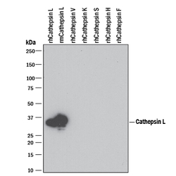
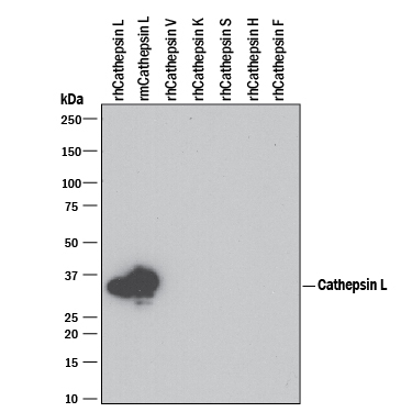
- Experimental details
- Detection of Recombinant Human and Mouse Cathepsin L by Western Blot. Western blot shows 100 ng of Recombinant Human Cathepsin L (Catalog # 952-CY), Recombinant Mouse Cathepsin L (Catalog # 1515-CY), Recombinant Human Cathepsin V (Catalog # 1080-CY), Recombinant Human Cathepsin K, Recombinant Human Cathepsin S (Catalog # 1183-CY), Recombinant Human Cathepsin H (Catalog # 7516-CY), and Recombinant Human Cathepsin F. PVDF Membrane was probed with 1 µg/mL of Rat Anti-Human/Mouse Cathepsin L Monoclonal Antibody (Catalog # MAB9521) followed by HRP-conjugated Anti-Rat IgG Secondary Antibody (Catalog # HAF005). A specific band was detected for Cathepsin L at approximately 35 kDa (as indicated). This experiment was conducted under reducing conditions and using Immunoblot Buffer Group 3. For natural samples, we recommend the use of Catalog # AF1515.
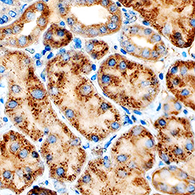
Supportive validation
- Submitted by
- Novus Biologicals (provider)
- Main image

- Experimental details
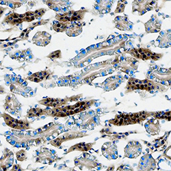
- Cathepsin L in Human Kidney. Cathepsin L was detected in immersion fixed paraffin-embedded sections of human kidney using Rat Anti-Human/Mouse Cathepsin L Monoclonal Antibody (Catalog # MAB9521) at 5 µg/mL overnight at 4 °C. Tissue was stained using the Anti-Rat HRP-DAB Cell & Tissue Staining Kit (brown; Catalog # CTS017) and counterstained with hematoxylin (blue). Specific staining was localized to cytoplasm in tubular epithelial cells. View our protocol for Chromogenic IHC Staining of Paraffin-embedded Tissue Sections.
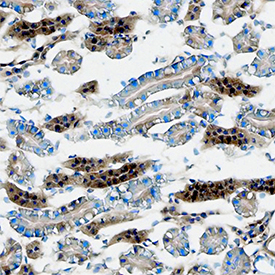
- Submitted by
- Novus Biologicals (provider)
- Main image
- Experimental details
- Cathepsin L in Mouse Kidney. Cathepsin L was detected in immersion fixed frozen sections of mouse kidney using Rat Anti-Human/Mouse Cathepsin L Monoclonal Antibody (Catalog # MAB9521) at 8 µg/mL overnight at 4 °C. Tissue was stained using the Anti-Rat HRP-DAB Cell & Tissue Staining Kit (brown; Catalog # CTS017) and counterstained with hematoxylin (blue). Specific staining was localized to cytoplasm in tubular epithelial cells. View our protocol for Chromogenic IHC Staining of Frozen Tissue Sections.