 Explore
 Validate
 Learn
Learn Western blot
 Immunoprecipitation
Antibody data
- Antibody Data
- Antigen structure
- References [7]
- Comments [0]
- Validations
- Western blot [1]
- Immunohistochemistry [2]
Submit
Validation data
Reference
Comment
Report error
- Product number
- MAB9521-100 - Provider product page

- Provider
- R&D Systems
- Product name
- Human/Mouse Cathepsin L Antibody
- Antibody type
- Monoclonal
- Description
- Protein A or G purified from hybridoma culture supernatant. Detects human and mouse Cathepsin L in direct ELISAs and Western blots. In direct ELISAs and Western blots, no cross-reactivity with recombinant human Cathepsin B, C, L2, O, S, or X/Z/P is observed. In Western blots, both the pro and active forms of recombinant human and mouse Cathspsin L are recognized.
- Reactivity
- Human, Mouse
- Host
- Rat
- Conjugate
- Unconjugated
- Antigen sequence
P07711- Isotype
- IgG
- Antibody clone number
- 204101
- Vial size
- 100 ug
- Storage
- Use a manual defrost freezer and avoid repeated freeze-thaw cycles. 12 months from date of receipt, -20 to -70 °C as supplied. 1 month, 2 to 8 °C under sterile conditions after reconstitution. 6 months, -20 to -70 °C under sterile conditions after reconstitution.
Submitted references A C9ORF72/SMCR8-containing complex regulates ULK1 and plays a dual role in autophagy.
CCT complex restricts neuropathogenic protein aggregation via autophagy.
Macrophages are crucial for epithelial cell death and adipocyte repopulation during mammary gland involution.
Stat3 controls lysosomal-mediated cell death in vivo.
The cystatin M/E-cathepsin L balance is essential for tissue homeostasis in epidermis, hair follicles, and cornea.
Primary tumour expression of the cysteine cathepsin inhibitor Stefin A inhibits distant metastasis in breast cancer.
Cystatin M/E is a high affinity inhibitor of cathepsin V and cathepsin L by a reactive site that is distinct from the legumain-binding site. A novel clue for the role of cystatin M/E in epidermal cornification.
Yang M, Liang C, Swaminathan K, Herrlinger S, Lai F, Shiekhattar R, Chen JF
Science advances 2016 Sep;2(9):e1601167
Science advances 2016 Sep;2(9):e1601167
CCT complex restricts neuropathogenic protein aggregation via autophagy.
Pavel M, Imarisio S, Menzies FM, Jimenez-Sanchez M, Siddiqi FH, Wu X, Renna M, O'Kane CJ, Crowther DC, Rubinsztein DC
Nature communications 2016 Dec 8;7:13821
Nature communications 2016 Dec 8;7:13821
Macrophages are crucial for epithelial cell death and adipocyte repopulation during mammary gland involution.
O'Brien J, Martinson H, Durand-Rougely C, Schedin P
Development (Cambridge, England) 2012 Jan;139(2):269-75
Development (Cambridge, England) 2012 Jan;139(2):269-75
Stat3 controls lysosomal-mediated cell death in vivo.
Kreuzaler PA, Staniszewska AD, Li W, Omidvar N, Kedjouar B, Turkson J, Poli V, Flavell RA, Clarkson RW, Watson CJ
Nature cell biology 2011 Mar;13(3):303-9
Nature cell biology 2011 Mar;13(3):303-9
The cystatin M/E-cathepsin L balance is essential for tissue homeostasis in epidermis, hair follicles, and cornea.
Zeeuwen PL, van Vlijmen-Willems IM, Cheng T, Rodijk-Olthuis D, Hitomi K, Hara-Nishimura I, John S, Smyth N, Reinheckel T, Hendriks WJ, Schalkwijk J
FASEB journal : official publication of the Federation of American Societies for Experimental Biology 2010 Oct;24(10):3744-55
FASEB journal : official publication of the Federation of American Societies for Experimental Biology 2010 Oct;24(10):3744-55
Primary tumour expression of the cysteine cathepsin inhibitor Stefin A inhibits distant metastasis in breast cancer.
Parker BS, Ciocca DR, Bidwell BN, Gago FE, Fanelli MA, George J, Slavin JL, Möller A, Steel R, Pouliot N, Eckhardt B, Henderson MA, Anderson RL
The Journal of pathology 2008 Feb;214(3):337-46
The Journal of pathology 2008 Feb;214(3):337-46
Cystatin M/E is a high affinity inhibitor of cathepsin V and cathepsin L by a reactive site that is distinct from the legumain-binding site. A novel clue for the role of cystatin M/E in epidermal cornification.
Cheng T, Hitomi K, van Vlijmen-Willems IM, de Jongh GJ, Yamamoto K, Nishi K, Watts C, Reinheckel T, Schalkwijk J, Zeeuwen PL
The Journal of biological chemistry 2006 Jun 9;281(23):15893-9
The Journal of biological chemistry 2006 Jun 9;281(23):15893-9
No comments: Submit comment
Supportive validation
- Submitted by
- R&D Systems (provider)
- Main image
- Experimental details
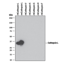
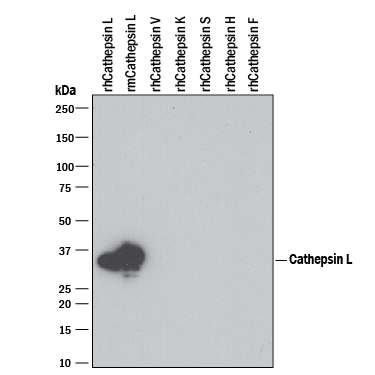
- Detection of Recombinant Human and Mouse Cathepsin L by Western Blot. Western blot shows 100 ng of Recombinant Human Cathepsin L (Catalog # 952-CY), Recombinant Mouse Cathepsin L (Catalog # 1515-CY), Recombinant Human Cathepsin V (Catalog # 1080-CY), Recombinant Human Cathepsin K, Recombinant Human Cathepsin S (Catalog # 1183-CY), Recombinant Human Cathepsin H (Catalog # 7516-CY), and Recombinant Human Cathepsin F. PVDF Membrane was probed with 1 µg/mL of Rat Anti-Human/Mouse Cathepsin L Monoclonal Antibody (Catalog # MAB9521) followed by HRP-conjugated Anti-Rat IgG Secondary Antibody (Catalog # HAF005). A specific band was detected for Cathepsin L at approximately 35 kDa (as indicated). This experiment was conducted under reducing conditions and using Immunoblot Buffer Group 3. For natural samples, we recommend the use of Catalog # AF1515.
Supportive validation
- Submitted by
- R&D Systems (provider)
- Main image
- Experimental details
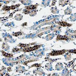
- Cathepsin L in Mouse Kidney. Cathepsin L was detected in immersion fixed frozen sections of mouse kidney using Rat Anti-Human/Mouse Cathepsin L Monoclonal Antibody (Catalog # MAB9521) at 8 µg/mL overnight at 4 °C. Tissue was stained using the Anti-Rat HRP-DAB Cell & Tissue Staining Kit (brown; Catalog # CTS017) and counterstained with hematoxylin (blue). Specific staining was localized to cytoplasm in tubular epithelial cells. View our protocol for Chromogenic IHC Staining of Frozen Tissue Sections.
- Submitted by
- R&D Systems (provider)
- Main image
- Experimental details
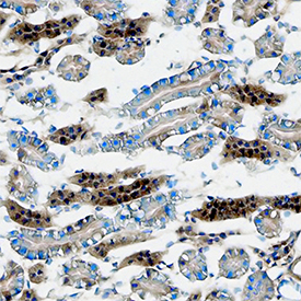
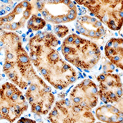
- Cathepsin L in Human Kidney. Cathepsin L was detected in immersion fixed paraffin-embedded sections of human kidney using Rat Anti-Human/Mouse Cathepsin L Monoclonal Antibody (Catalog # MAB9521) at 5 µg/mL overnight at 4 °C. Tissue was stained using the Anti-Rat HRP-DAB Cell & Tissue Staining Kit (brown; Catalog # CTS017) and counterstained with hematoxylin (blue). Specific staining was localized to cytoplasm in tubular epithelial cells. View our protocol for Chromogenic IHC Staining of Paraffin-embedded Tissue Sections.
