 Explore
 Validate
 Learn
Learn Western blot
 ELISA
ELISAAntibody data
- Antibody Data
- Antigen structure
- References [0]
- Comments [0]
- Validations
- Western blot [2]
- Immunohistochemistry [1]
Submit
Validation data
Reference
Comment
Report error
- Product number
- PA5-47971 - Provider product page

- Provider
- Invitrogen Antibodies
- Product name
- Cathepsin L Polyclonal Antibody
- Antibody type
- Polyclonal
- Antigen
- Recombinant full-length protein
- Description
- In sandwich ELISAs, less than 0.2% cross-reactivity with recombinant mouse Cathepsin L, recombinant human (rh) Cathepsin A, rhCathepsin B, rhCathepsin C, rhCathepsin D, rhCathepsin E, rhCathepsin S, and rhCathepsin V is observed. Reconstitute at 0.2 mg/mL in sterile PBS. Endoxin level is
- Reactivity
- Human
- Host
- Goat
- Isotype
- IgG
- Vial size
- 100 µg
- Concentration
- 0.2 mg/mL
- Storage
- -20° C, Avoid Freeze/Thaw Cycles
No comments: Submit comment
Supportive validation
- Submitted by
- Invitrogen Antibodies (provider)
- Main image

- Experimental details
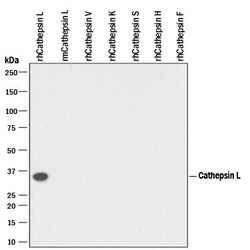
- Western blot analysis of Cathepsin L in 10 ng recombinant Human Cathepsin L, recombinant Mouse Cathepsin L, recombinant Human Cathepsin V, recombinant Human Cathepsin K, recombinant Human Cathepsin S, recombinant Human Cathepsin H, and recombinant Human Cathepsin F. Samples were incubated in Cathepsin L polyclonal antibody (Product # PA5-47971) using a dilution of 0.1 µg/mL followed by a HRP-conjugated Anti-Goat IgG secondary antibody. A specific band was detected for Cathepsin L at approximately 35 kDa (as indicated). This experiment was conducted under reducing conditions.

- Submitted by
- Invitrogen Antibodies (provider)
- Main image
- Experimental details
- Western blot analysis of Cathepsin L in 10 ng recombinant Human Cathepsin L, recombinant Mouse Cathepsin L, recombinant Human Cathepsin V, recombinant Human Cathepsin K, recombinant Human Cathepsin S, recombinant Human Cathepsin H, and recombinant Human Cathepsin F. Samples were incubated in Cathepsin L polyclonal antibody (Product # PA5-47971) using a dilution of 0.1 µg/mL followed by a HRP-conjugated Anti-Goat IgG secondary antibody. A specific band was detected for Cathepsin L at approximately 35 kDa (as indicated). This experiment was conducted under reducing conditions.

Supportive validation
- Submitted by
- Invitrogen Antibodies (provider)
- Main image
- Experimental details
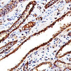
- Immunohistochemical analysis of Cathepsin L in immersion fixed paraffin-embedded sections of human kidney. Samples were incubated in Cathepsin L polyclonal antibody (Product # PA5-47971) using a dilution of 15 µg/mL overnight at 4 °C. Tissue was stained using the Anti-Goat HRP-DAB Cell & Tissue Staining Kit (brown) and counterstained with hematoxylin (blue). Specific staining was localized to convoluted tubules.
