Explore
 Validate
 Learn
Learn Western blot
 Immunoprecipitation
Antibody data
- Antibody Data
- Antigen structure
- References [2]
- Comments [0]
- Validations
- Western blot [3]
Submit
Validation data
Reference
Comment
Report error
- Product number
- AF1408 - Provider product page

- Provider
- R&D Systems
- Product name
- Human/Rat Cystatin B Antibody
- Antibody type
- Polyclonal
- Description
- Antigen Affinity-purified. Detects human and rat Cystatin B in Western blots. In Western blots, less than 2% cross-reactivity with recombinant human (rh) Cystatin A, recombinant mouse Cystatin B, rhCystatin C, rhCystatin D, rhCystatin E/M, rhCystatin S, rhCystatin SA, and rhCystatin SN is observed.
- Reactivity
- Human, Rat
- Host
- Goat
- Conjugate
- Unconjugated
- Antigen sequence
P04080- Isotype
- IgG
- Vial size
- 100 ug
- Concentration
- LYOPH
- Storage
- Use a manual defrost freezer and avoid repeated freeze-thaw cycles. 12 months from date of receipt, -20 to -70 °C as supplied. 1 month, 2 to 8 °C under sterile conditions after reconstitution. 6 months, -20 to -70 °C under sterile conditions after reconstitution.
Submitted references Modulation of Receptor Protein Tyrosine Phosphatase Sigma Increases Chondroitin Sulfate Proteoglycan Degradation through Cathepsin B Secretion to Enhance Axon Outgrowth.
Regulation of TGF-β1-driven differentiation of human lung fibroblasts: emerging roles of cathepsin B and cystatin C.
Tran AP, Sundar S, Yu M, Lang BT, Silver J
The Journal of neuroscience : the official journal of the Society for Neuroscience 2018 Jun 6;38(23):5399-5414
The Journal of neuroscience : the official journal of the Society for Neuroscience 2018 Jun 6;38(23):5399-5414
Regulation of TGF-β1-driven differentiation of human lung fibroblasts: emerging roles of cathepsin B and cystatin C.
Kasabova M, Joulin-Giet A, Lecaille F, Gilmore BF, Marchand-Adam S, Saidi A, Lalmanach G
The Journal of biological chemistry 2014 Jun 6;289(23):16239-51
The Journal of biological chemistry 2014 Jun 6;289(23):16239-51
No comments: Submit comment
Supportive validation
- Submitted by
- R&D Systems (provider)
- Main image
- Experimental details
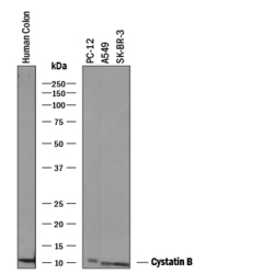
- Detection of Human and Rat Cystatin B by Western Blot. Western blot shows lysates of human colon tissue, PC-12 rat adrenal pheochromocytoma cell line, A549 human lung carcinoma cell line, and SK-BR-3 human breast cancer cell line. PVDF membrane was probed with 1 µg/mL of Goat Anti-Human/Rat Cystatin B Antigen Affinity-purified Polyclonal Antibody (Catalog # AF1408) followed by HRP-conjugated Anti-Goat IgG Secondary Antibody (Catalog # HAF019). A specific band was detected for Cystatin B at approximately 11 kDa (as indicated). This experiment was conducted under reducing conditions and using Immunoblot Buffer Group 1.

- Submitted by
- R&D Systems (provider)
- Main image
- Experimental details
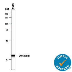
- Detection of Human Cystatin B by Simple Western<SUP abp="263">TM. Simple Western lane view shows lysates of A549 human lung carcinoma cell line, loaded at 0.2 mg/mL. A specific band was detected for Cystatin B at approximately 18 kDa (as indicated) using 10 µg/mL of Goat Anti-Human/Rat Cystatin B Antigen Affinity-purified Polyclonal Antibody (Catalog # AF1408) followed by 1:50 dilution of HRP-conjugated Anti-Goat IgG Secondary Antibody (Catalog # HAF109). This experiment was conducted under reducing conditions and using the 12-230 kDa separation system.

- Submitted by
- R&D Systems (provider)
- Main image

- Experimental details
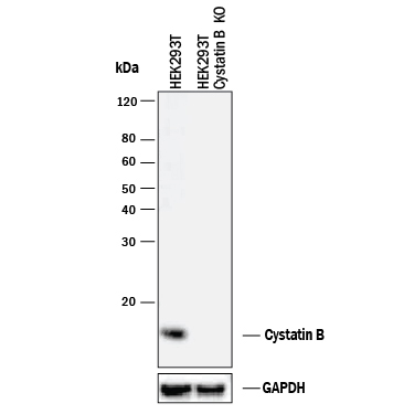
- Western Blot Shows Human Cystatin B Specificity by Using Knockout Cell Line. Western blot shows lysates of HEK293T human embryonic kidney parental cell line and Cystatin B knockout HEK293T cell line (KO). PVDF membrane was probed with 1 µg/mL of Goat Anti-Human/Rat Cystatin B Antigen Affinity-purified Polyclonal Antibody (Catalog # AF1408) followed by HRP-conjugated Anti-Goat IgG Secondary Antibody (Catalog # HAF017). A specific band was detected for Cystatin B at approximately 12 kDa (as indicated) in the parental HEK293T cell line, but is not detectable in knockout HEK293T cell line. GAPDH (Catalog # AF5718) is shown as a loading control. This experiment was conducted under reducing conditions and using Immunoblot Buffer Group 1.