 Explore
 Validate
 Learn
Learn Western blot
 ELISA
ELISAAntibody data
- Antibody Data
- Antigen structure
- References [0]
- Comments [0]
- Validations
- Western blot [2]
- Immunocytochemistry [1]
- Immunoprecipitation [1]
- Other assay [1]
Submit
Validation data
Reference
Comment
Report error
- Product number
- PA5-95926 - Provider product page

- Provider
- Invitrogen Antibodies
- Product name
- AKR1C3 Polyclonal Antibody
- Antibody type
- Polyclonal
- Antigen
- Recombinant full-length protein
- Description
- Immunogen sequence: MDSKHQCVKL NDGHFMPVLG FGTYAPPEVP RSKALEVTKL AIEAGFRHID SAHLYNNEEQ VGLAIRSKIA DGSVKREDIF YTSKLWSTFH RPELVRPALE NSLKKAQLDY VDLYLIHSPM SLKPGEELSP TDENGKVIFD IVDLCTTWEA MEKCKDAGLA KSIGVSNFNR RQLEMILNKP GLKYKPVCNQ VECHPYFNRS KLLDFCKSKD IVLVAYSALG SQRDKRWVDP NSPVLLEDPV LCALAKKHKR TPALIALRYQ LQRGVVVLAK SYNEQRIRQN VQVFEFQLTA EDMKAIDGLD RNLHYFNSDS FASHPNYPYS DEY; Positive Samples: U-87MG, 293T, LO2, A-549, DU145, Mouse kidney; Cellular Location: Cytoplasm
- Reactivity
- Human, Mouse
- Host
- Rabbit
- Isotype
- IgG
- Vial size
- 100 μL
- Concentration
- 0.6 mg/mL
- Storage
- -20°C, Avoid Freeze/Thaw Cycles
No comments: Submit comment
Supportive validation
- Submitted by
- Invitrogen Antibodies (provider)
- Main image

- Experimental details
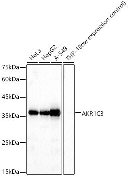
- Western blot analysis of AKR1C3 in various lysates. Samples were incubated with AKR1C3 Polyclonal antibody (Product # PA5-95926) using a dilution of 1:1,000, followed by HRP Goat Anti-Rabbit IgG (H+L) at a dilution of 1:10,000. Lysates/proteins: 25 µg per lane. Blocking buffer: 3% nonfat dry milk in TBST. Detection: ECL Basic Kit. Exposure time: 10s.
- Submitted by
- Invitrogen Antibodies (provider)
- Main image

- Experimental details
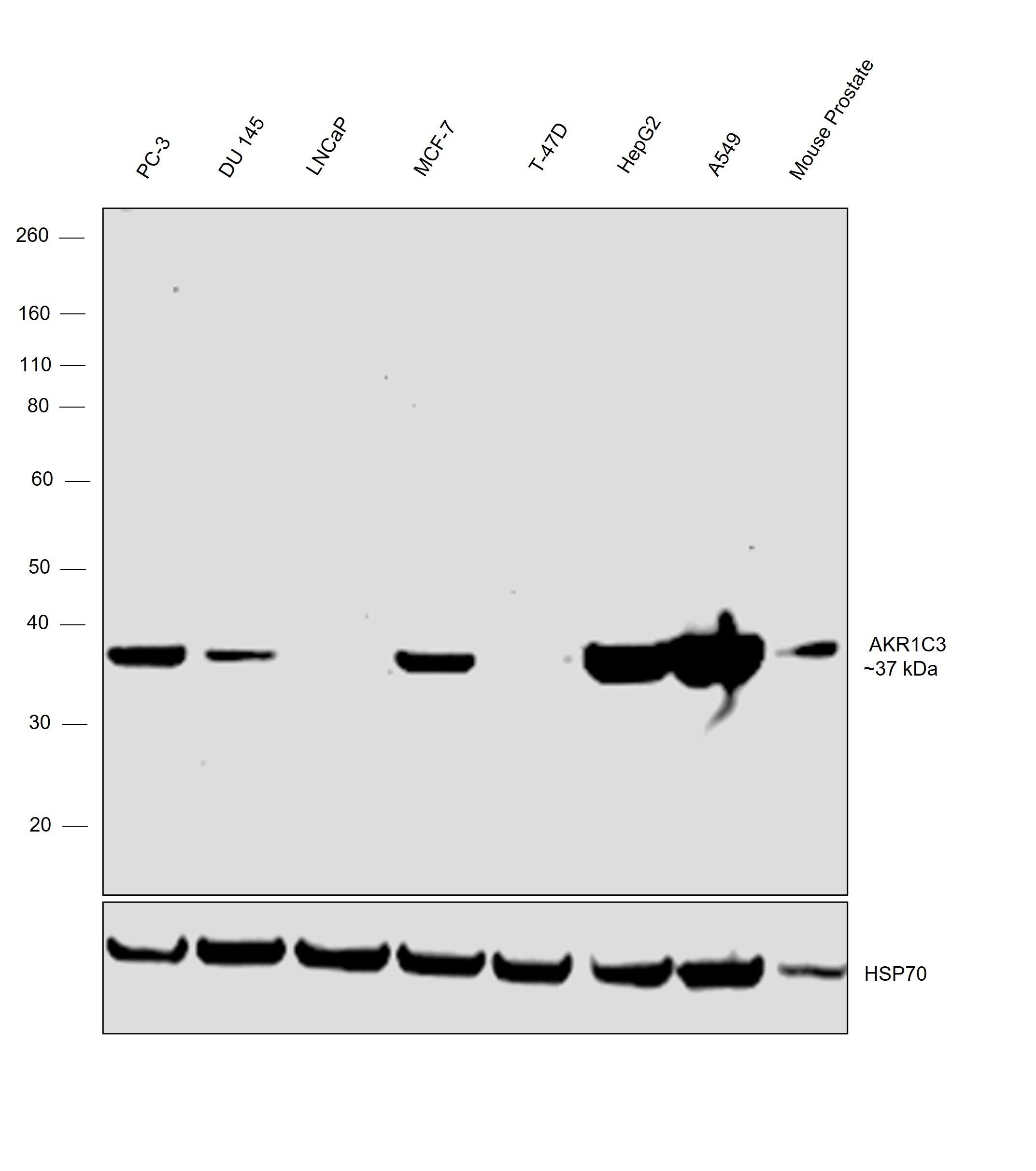
- Western blot was performed using Anti-AKR1C3 Polyclonal Antibody (Product # PA5-95926) and a 37 kDa band corresponding to 17-beta-HSD 5; AKR1C3 was observed across the panel tested except for LNCaP and T-47D which are reported to be low expressors of AKR1C3. Whole cell extracts (30 µg lysate) of PC-3 (Lane 1), DU 145 (Lane 2), LNCaP (Lane 3), MCF7 (Lane 4), T-47D (Lane 5), Hep G2 (Lane 6), A549 (Lane 7), Mouse Prostate (Lane 8) were electrophoresed using NuPAGE™ 4-12% Bis-Tris Protein Gel (Product # NP0321BOX), 10 well. Resolved proteins were then transferred onto a nitrocellulose membrane (Product # IB23001) by iBlot® 2 Dry Blotting System (Product # IB21001). The blot was probed with the primary antibody (1:1000 dilution) and detected by chemiluminescence with Goat anti-Rabbit IgG (Heavy Chain) Superclonal™ Recombinant Secondary Antibody, HRP (Product # A27036, 1:20,000 dilution) using the iBright™ FL1500 Imaging System (Product # A44115). Chemiluminescent detection was performed using SuperSignal™ West Pico PLUS Chemiluminescent Substrate (Product # 34580).
Supportive validation
- Submitted by
- Invitrogen Antibodies (provider)
- Main image

- Experimental details
- Immunofluorescence analysis of AKR1C3 in NIH/3T3 cells. Samples were incubated with AKR1C3 Polyclonal antibody (Product # PA5-95926) using a dilution of 1:100. Blue: DAPI for nuclear staining.

Supportive validation
- Submitted by
- Invitrogen Antibodies (provider)
- Main image
- Experimental details
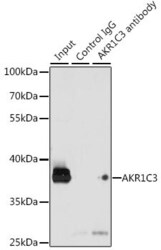
- Immunoprecipitation of AKR1C3 in 200 μg extracts of K-562 cells. Samples were precipitated with 3 μg AKR1C3 Polyclonal antibody (Product # PA5-95926). Western blot was performed from the immunoprecipitate using AKR1C3 Polyclonal antibody (Product # PA5-95926) at a dilution of 1:1,000.

Supportive validation
- Submitted by
- Invitrogen Antibodies (provider)
- Main image

- Experimental details
- Immunoprecipitation analysis of AKR1C3 was performed in 200 µg extracts of K-562 cells using AKR1C3 Polyclonal Antibody (Product # PA5-95926). Western blot was performed from the immunoprecipitate using AKR1C3 Polyclonal Antibody.
