 Explore
 Validate
 Learn
LearnPA5-106891
antibody from Invitrogen Antibodies
Targeting: AKR1C3
DDX, HAKRB, HSD17B5, KIAA0119, PGFS
 Western blot
Antibody data
- Antibody Data
- Antigen structure
- References [0]
- Comments [0]
- Validations
- Western blot [2]
- Immunocytochemistry [3]
- Immunohistochemistry [1]
Submit
Validation data
Reference
Comment
Report error
- Product number
- PA5-106891 - Provider product page

- Provider
- Invitrogen Antibodies
- Product name
- AKR1C3 Polyclonal Antibody
- Antibody type
- Polyclonal
- Antigen
- Synthetic peptide
- Description
- Antibody detects endogenous levels of total AKR1C3.
- Reactivity
- Human, Mouse, Rat
- Host
- Rabbit
- Isotype
- IgG
- Vial size
- 100 µL
- Concentration
- 1 mg/mL
- Storage
- -20°C
No comments: Submit comment
Supportive validation
- Submitted by
- Invitrogen Antibodies (provider)
- Main image

- Experimental details
- Western blot analysis of AKR1C3 in mouse brain. Samples were incubated with AKR1C3 polyclonal antibody (Product # PA5-106891).

- Submitted by
- Invitrogen Antibodies (provider)
- Main image

- Experimental details
- Western blot was performed using Anti-AKR1C3 Polyclonal Antibody (Product # PA5-106891) and a 37 kDa band corresponding to 17-beta-HSD 5; AKR1C3 was observed across the panel tested except for LNCaP and T-47D which are reported to be low expressors of AKR1C3. Whole cell extracts (30 µg lysate) of PC-3 (Lane 1), DU 145 (Lane 2), LNCaP (Lane 3), MCF7 (Lane 4), T-47D (Lane 5), Hep G2 (Lane 6), A549 (Lane 7), Mouse Prostate (Lane 8), Mouse Colon (Lane 9) were electrophoresed using NuPAGE™ 4-12% Bis-Tris Protein Gel (Product # NP0322BOX), 12 well. Resolved proteins were then transferred onto a nitrocellulose membrane (Product # IB23001) by iBlot® 2 Dry Blotting System (Product # IB21001). The blot was probed with the primary antibody (1:1000 dilution) and detected by chemiluminescence with Goat anti-Rabbit IgG (H+L) Superclonal™ Recombinant Secondary Antibody, HRP (Product # A27036, 1:20,000 dilution) using the iBright™ FL1500 Imaging System (Product # A44115). Chemiluminescent detection was performed using SuperSignal™ West Pico PLUS Chemiluminescent Substrate (Product # 34580).

Supportive validation
- Submitted by
- Invitrogen Antibodies (provider)
- Main image

- Experimental details
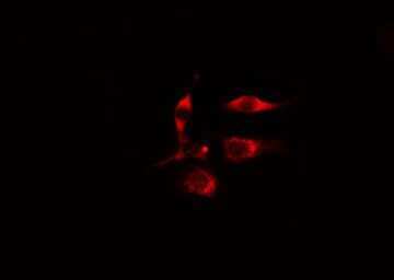
- Immunofluorescent analysis of AKR1C3 in HepG2 cell lysate. Samples were fixed with paraformaldehyde, permeabilized with 0.1% Triton X-100, blocked with 10% serum (45 min at 25°C) incubated with AKR1C3 polyclonal antibody (Product # PA5-106891) using a dilution of 1:200 (1 hr, 37°C), and followed by goat anti-rabbit IgG Alexa Fluor 594 at a dilution of 1:600.

- Submitted by
- Invitrogen Antibodies (provider)
- Main image

- Experimental details
- Immunofluorescent analysis of AKR1C3 in HeLa cells. Samples were fixed with paraformaldehyde, permeabilized with 0.1% Triton X-100, blocked with 10% serum (45 min at 25°C) incubated with AKR1C3 polyclonal antibody (Product # PA5-106891) using a dilution of 1:200 (1 hr, 37°C), and followed by goat anti-rabbit IgG Alexa Fluor 594 at a dilution of 1:600.
- Submitted by
- Invitrogen Antibodies (provider)
- Main image

- Experimental details
- Immunofluorescent analysis of AKR1C3 in HepG2 cell lysate. Samples were fixed with paraformaldehyde, permeabilized with 0.1% Triton X-100, blocked with 10% serum (45 min at 25°C) incubated with AKR1C3 polyclonal antibody (Product # PA5-106891) using a dilution of 1:200 (1 hr, 37°C), and followed by goat anti-rabbit IgG Alexa Fluor 594 at a dilution of 1:600.

Supportive validation
- Submitted by
- Invitrogen Antibodies (provider)
- Main image

- Experimental details
- Immunohistochemistry analysis of paraffin-embedded AKR1C3 in mouse spleen tissue. Antigen retrieval was performed using citrate buffer. Samples were blocked with blocking buffer (1.5 hr, 22°C), incubated with AKR1C3 polyclonal antibody (Product # PA5-106891) using a dilution of 1:100 (1.5 hr, 22°C), followed by HRP conjugated goat anti-rabbit.
