Explore
 Validate
 Learn
LearnMHCXCR404
antibody from Invitrogen Antibodies
Targeting: CXCR4
CD184, D2S201E, fusin, HM89, HSY3RR, LESTR, NPY3R, NPYR, NPYY3R
 Flow cytometry
 Other assay
Antibody data
- Antibody Data
- Antigen structure
- References [11]
- Comments [0]
- Validations
- Other assay [4]
Submit
Validation data
Reference
Comment
Report error
- Product number
- MHCXCR404 - Provider product page

- Provider
- Invitrogen Antibodies
- Product name
- CXCR4 Monoclonal Antibody (12G5), PE
- Antibody type
- Monoclonal
- Antigen
- Other
- Description
- R-phycoerythrin (PE) is a stable and highly soluble phycobiliprotein which provides maximal absorbance and fluorescence without susceptibility to internal or external fluorescence quenching, thus providing an exceptional quantum yields and molar extinction coefficients.
- Reactivity
- Human
- Host
- Mouse
- Conjugate
- Yellow dye
- Isotype
- IgG
- Antibody clone number
- 12G5
- Vial size
- 500 µL
- Storage
- 4° C, store in dark
Submitted references Functional Genomic Screening in Human Pluripotent Stem Cells Reveals New Roadblocks in Early Pancreatic Endoderm Formation.
CDKN2A-Mutated Pancreatic Ductal Organoids from Induced Pluripotent Stem Cells to Model a Cancer Predisposition Syndrome.
Transcriptional changes and the role of ONECUT1 in hPSC pancreatic differentiation.
Modeling plasticity and dysplasia of pancreatic ductal organoids derived from human pluripotent stem cells.
Differentiation of human pluripotent stem cells into pancreatic duct-like organoids.
Clampdown of inflammation in aging and anticancer therapies by limiting upregulation and activation of GPCR, CXCR4.
Genome Editing of the CYP1A1 Locus in iPSCs as a Platform to Map AHR Expression throughout Human Development.
Multisystemic Disease Modeling of Liver-Derived Protein Folding Disorders Using Induced Pluripotent Stem Cells (iPSCs).
The in vitro generation of lung and airway progenitor cells from human pluripotent stem cells.
KPNA2 expression is an independent adverse predictor of biochemical recurrence after radical prostatectomy.
Co-culture of hematopoietic stem cells with mesenchymal stem cells increases VCAM-1-dependent migration of primitive hematopoietic stem cells.
Krüger J, Breunig M, Pasquini LP, Morawe M, Groß A, Arnold F, Russell R, Seufferlein T, Azoitei N, Kestler HA, Julier C, Heller S, Hohwieler M, Kleger A
Cells 2022 Feb 8;11(3)
Cells 2022 Feb 8;11(3)
CDKN2A-Mutated Pancreatic Ductal Organoids from Induced Pluripotent Stem Cells to Model a Cancer Predisposition Syndrome.
Merkle J, Breunig M, Schmid M, Allgöwer C, Krüger J, Melzer MK, Bens S, Siebert R, Perkhofer L, Azoitei N, Seufferlein T, Heller S, Meier M, Müller M, Kleger A, Hohwieler M
Cancers 2021 Oct 13;13(20)
Cancers 2021 Oct 13;13(20)
Transcriptional changes and the role of ONECUT1 in hPSC pancreatic differentiation.
Heller S, Li Z, Lin Q, Geusz R, Breunig M, Hohwieler M, Zhang X, Nair GG, Seufferlein T, Hebrok M, Sander M, Julier C, Kleger A, Costa IG
Communications biology 2021 Nov 17;4(1):1298
Communications biology 2021 Nov 17;4(1):1298
Modeling plasticity and dysplasia of pancreatic ductal organoids derived from human pluripotent stem cells.
Breunig M, Merkle J, Wagner M, Melzer MK, Barth TFE, Engleitner T, Krumm J, Wiedenmann S, Cohrs CM, Perkhofer L, Jain G, Krüger J, Hermann PC, Schmid M, Madácsy T, Varga Á, Griger J, Azoitei N, Müller M, Wessely O, Robey PG, Heller S, Dantes Z, Reichert M, Günes C, Bolenz C, Kuhn F, Maléth J, Speier S, Liebau S, Sipos B, Kuster B, Seufferlein T, Rad R, Meier M, Hohwieler M, Kleger A
Cell stem cell 2021 Jun 3;28(6):1105-1124.e19
Cell stem cell 2021 Jun 3;28(6):1105-1124.e19
Differentiation of human pluripotent stem cells into pancreatic duct-like organoids.
Breunig M, Merkle J, Melzer MK, Heller S, Seufferlein T, Meier M, Hohwieler M, Kleger A
STAR protocols 2021 Dec 17;2(4):100913
STAR protocols 2021 Dec 17;2(4):100913
Clampdown of inflammation in aging and anticancer therapies by limiting upregulation and activation of GPCR, CXCR4.
Nair RR, Madiwale SV, Saini DK
NPJ aging and mechanisms of disease 2018;4:9
NPJ aging and mechanisms of disease 2018;4:9
Genome Editing of the CYP1A1 Locus in iPSCs as a Platform to Map AHR Expression throughout Human Development.
Smith BW, Stanford EA, Sherr DH, Murphy GJ
Stem cells international 2016;2016:2574152
Stem cells international 2016;2016:2574152
Multisystemic Disease Modeling of Liver-Derived Protein Folding Disorders Using Induced Pluripotent Stem Cells (iPSCs).
Leung A, Murphy GJ
Methods in molecular biology (Clifton, N.J.) 2016;1353:261-70
Methods in molecular biology (Clifton, N.J.) 2016;1353:261-70
The in vitro generation of lung and airway progenitor cells from human pluripotent stem cells.
Huang SX, Green MD, de Carvalho AT, Mumau M, Chen YW, D'Souza SL, Snoeck HW
Nature protocols 2015 Mar;10(3):413-25
Nature protocols 2015 Mar;10(3):413-25
KPNA2 expression is an independent adverse predictor of biochemical recurrence after radical prostatectomy.
Mortezavi A, Hermanns T, Seifert HH, Baumgartner MK, Provenzano M, Sulser T, Burger M, Montani M, Ikenberg K, Hofstädter F, Hartmann A, Jaggi R, Moch H, Kristiansen G, Wild PJ
Clinical cancer research : an official journal of the American Association for Cancer Research 2011 Mar 1;17(5):1111-21
Clinical cancer research : an official journal of the American Association for Cancer Research 2011 Mar 1;17(5):1111-21
Co-culture of hematopoietic stem cells with mesenchymal stem cells increases VCAM-1-dependent migration of primitive hematopoietic stem cells.
Perdomo-Arciniegas AM, Vernot JP
International journal of hematology 2011 Dec;94(6):525-32
International journal of hematology 2011 Dec;94(6):525-32
No comments: Submit comment
Supportive validation
- Submitted by
- Invitrogen Antibodies (provider)
- Main image
- Experimental details
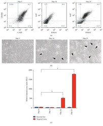
- Figure 2 Hepatocyte specification yields luciferase expression in cells derived from CYP1A1 reporter iPSCs. (a) iPSCs were differentiated towards CXCR4+/C-KIT+ definitive endoderm (day 5) followed by FOXA1+/AAT+ hepatic progenitors (day 14) that grew in number and were the majority of the culture by day 25. (b) Micrographs show homogenous morphology of definitive endoderm cultures (day 5), but by day 14, hepatic-like cells begin to emerge (denoted by black arrowheads) and are observed more frequently by day 25. (c) Concomitant with hepatic specification, luciferase levels significantly increase ( N = 3, * P < 0.0005, Student's t -test).
- Conjugate
- Yellow dye
- Submitted by
- Invitrogen Antibodies (provider)
- Main image
- Experimental details
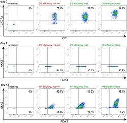
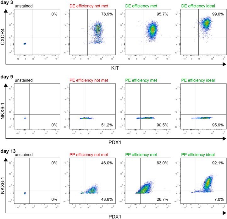
- Figure 3 Quality check via flow cytometry Differentiation efficiencies at definitive endoderm (DE, d3), pancreatic endoderm (PE, d9), and pancreatic progenitor (PP, d13) stage are measured by flow cytometry. Our quality standards are 95% CXCR4 + /KIT + cells at DE stage, 90% PDX1 + cells at PE stage, and 60% PDX1 + /NKX6-1 + cells at PP stage.
- Conjugate
- Yellow dye
- Submitted by
- Invitrogen Antibodies (provider)
- Main image
- Experimental details
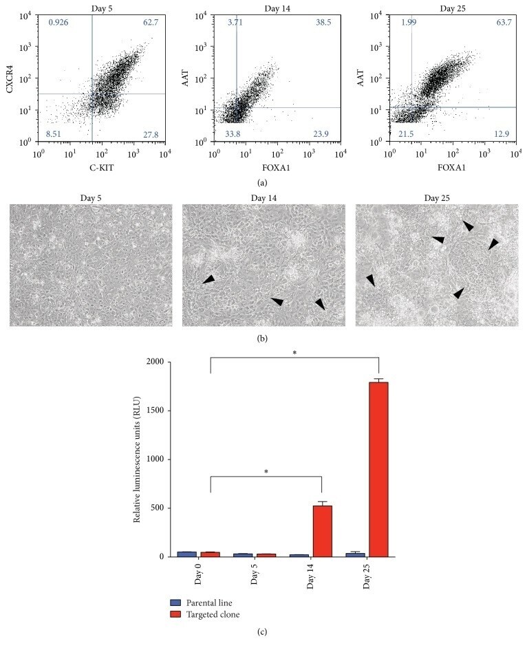
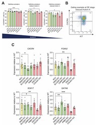
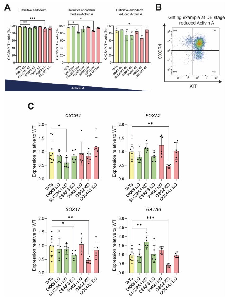
- Figure 4 DSC2 and SLC22A1 KO lines show an impaired differentiation into definitive endoderm. ( A ) Newly generated KO cell lines were differentiated towards DE stage with either the standard induction protocol (left), or with only 10% or 5% (right) of standard activin A concentration. ( B ) Differentiation efficiency was analysed by flow cytometry of DE markers CXCR4 and KIT, as shown in a representative FACS plot. ( C ) From cells generated under optimal culture conditions, RNA samples were taken and analysed via qPCR for expression levels of DE markers CXCR4 , FOXA2 , SOX17, and GATA6 . Experiments with standard culture conditions were performed 3 times, using 2 different clones per genotype in technical duplicates (dots represent means of duplicates). Experiments with reduced levels of activin A were performed once with 2 different clones per genotype and in duplicates (dots represent means of these duplicates). Gene expression was first normalized to housekeeping gene HMBS and then normalized to WT gene expression. Mann-Whitney test was used for analysis; error bars represent mean +- SD, *** p < 0.001, ** p < 0.01, * p < 0.05.
- Conjugate
- Yellow dye
- Submitted by
- Invitrogen Antibodies (provider)
- Main image
- Experimental details
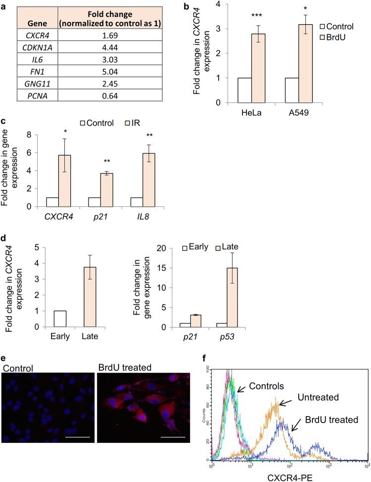
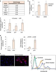
- Fig. 1 CXCR4 expression analysis in response to DNA damage. a Gene expression changes after BrdU treatment in HeLa cells. Expression pattern of genes as indicated after 48 h of BrdU treatment (100 muM) by microarray analysis. Data extracted from the microarray experiment is reported in Supplementary Fig. S1a ( n = 2). The numbers indicate fold change in gene expression during BrdU treatment wrt Control (normalized as 1). b CXCR4 gene expression analysis after DNA damage. HeLa and A549 cells were treated for 48 h with 100 muM BrdU followed by qRT-PCR analysis of CXCR4 expression. The values were normalized to beta-actin expression and then wrt control cells to calculate fold changes. Results shown are mean +- s.e.m. * p 3). d Gene expression analysis during replicative exhaustion mediated DNA damage and senescence. Expression analysis of CXCR4 (left) and DDR genes P21 and P53 (right) in early (20 PDL) and late (40 PDL) passage MRC5 cells by qRT-PCR. The values were normalized to beta-actin expression and then wrt control cells to calculate fold changes ( n
- Conjugate
- Yellow dye