 Explore
 Validate
 Learn
Learn711271
antibody from Invitrogen Antibodies
Targeting: CXCR4
CD184, D2S201E, fusin, HM89, HSY3RR, LESTR, NPY3R, NPYR, NPYY3R
 Western blot
Antibody data
- Antibody Data
- Antigen structure
- References [0]
- Comments [0]
- Validations
- Western blot [2]
- Immunocytochemistry [1]
Submit
Validation data
Reference
Comment
Report error
- Product number
- 711271 - Provider product page

- Provider
- Invitrogen Antibodies
- Product name
- Phospho-CXCR4 (Ser324, Ser325) Recombinant Polyclonal Antibody (4 HCLC)
- Antibody type
- Polyclonal
- Antigen
- Synthetic peptide
- Reactivity
- Human
- Host
- Rabbit
- Isotype
- IgG
- Antibody clone number
- 4 HCLC
- Vial size
- 100 µg
- Concentration
- 0.5 mg/mL
- Storage
- Store at 4°C short term. For long term storage, store at -20°C, avoiding freeze/thaw cycles.
No comments: Submit comment
Supportive validation
- Submitted by
- Invitrogen Antibodies (provider)
- Main image
- Experimental details
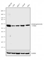
- Western blot analysis was performed on Membrane enriched extracts (30 µg lysate) of HEK-293 (Lane 1), A549 (Lane 2), MCF-7 (Lane 3), LNCaP (Lane 4) and HeLa (Lane 5). The blots were probed with Anti-CXCR4 [pS324/325] Recombinant Rabbit Polyclonal Antibody (Product # 711271, 2.5 µg/mL) and detected by chemiluminescence using Goat anti-Rabbit IgG (H+L) Superclonal™ Secondary Antibody, HRP conjugate (Product # A27036, 0.25 µg/mL, 1:4000 dilution). A 55 kDa band corresponding to CXCR4 [pS324/425] was observed across the cell lines tested. Known quantity of protein samples were electrophoresed using Novex®NuPAGE®4-12% Bis-Tris gel (Product # NP0322BOX), XCell SureLock™ Electrophoresis System (Product # EI0002) and Novex® Sharp Pre-Stained Protein Standard (Product # LC5800). Resolved proteins were then transferred onto a nitrocellulose membrane with iBlot® Dry Blotting System (Product # IB21001). The membrane was probed with the relevant primary and secondary Antibody following blocking with 5% skimmed milk. Chemiluminescent detection was performed using Pierce™ ECL Western blotting Substrate (Product # 32106).

- Submitted by
- Invitrogen Antibodies (provider)
- Main image
- Experimental details
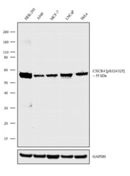
- Western blot analysis was performed on Membrane enriched extracts (30 µg lysate) of HEK-293 (Lane 1), A549 (Lane 2), MCF-7 (Lane 3), LNCaP (Lane 4) and HeLa (Lane 5). The blots were probed with Anti-CXCR4 [pS324/325] Recombinant Rabbit Polyclonal Antibody (Product # 711271, 2.5 µg/mL) and detected by chemiluminescence using Goat anti-Rabbit IgG (H+L) Superclonal™ Secondary Antibody, HRP conjugate (Product # A27036, 0.25 µg/mL, 1:4000 dilution). A 55 kDa band corresponding to CXCR4 [pS324/425] was observed across the cell lines tested. Known quantity of protein samples were electrophoresed using Novex®NuPAGE®4-12% Bis-Tris gel (Product # NP0322BOX), XCell SureLock™ Electrophoresis System (Product # EI0002) and Novex® Sharp Pre-Stained Protein Standard (Product # LC5800). Resolved proteins were then transferred onto a nitrocellulose membrane with iBlot® Dry Blotting System (Product # IB21001). The membrane was probed with the relevant primary and secondary Antibody following blocking with 5% skimmed milk. Chemiluminescent detection was performed using Pierce™ ECL Western blotting Substrate (Product # 32106).

Supportive validation
- Submitted by
- Invitrogen Antibodies (provider)
- Main image
- Experimental details
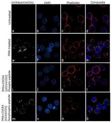
- For immunofluorescence analysis Jurkat cells were fixed and permeabilized for detection of endogenous CXCR4 [pS324/325] using Anti- CXCR4 [pS324/325] Recombinant Rabbit Polyclonal Antibody (Product # 711271, 5 µg/mL) and labeled with Goat anti-Rabbit IgG (H+L) Superclonal™ Secondary Antibody, Alexa Fluor® 488 conjugate (Product # A27034, 1:2000). Nuclei (blue) were stained using SlowFade® Gold Antifade Mountant with DAPI (Product # S36938), and Rhodamine Phalloidin (Product # R415, 1:300) was used for cytoskeletal F-actin (red) staining. Detection and localization of CXCR4 [pS324/325] (green)on the membrane can be clearly observed in cells treated with PMA (40 ng/ML, 30 min) as compared to untreated cells. Antibody specificity was demonstrated by competition with the CXCR4 [pS324/325] phospho peptide, which results in loss of signal. No competition was observed with the non-phospho peptide. The images were captured at 60X magnification.
