Explore
 Validate
 Learn
LearnNBP1-77067
antibody from Novus Biologicals
Targeting: CXCR4
CD184, D2S201E, fusin, HM89, HSY3RR, LESTR, NPY3R, NPYR, NPYY3R
 Western blot
 ELISA
ELISAAntibody data
- Antibody Data
- Antigen structure
- References [4]
- Comments [0]
- Validations
- Western blot [7]
- Immunohistochemistry [1]
- Flow cytometry [2]
Submit
Validation data
Reference
Comment
Report error
- Product number
- NBP1-77067 - Provider product page

- Provider
- Novus Biologicals
- Proper citation
- Novus Cat#NBP1-77067, RRID:AB_11005253
- Product name
- Rabbit Polyclonal CXCR4 Antibody
- Antibody type
- Polyclonal
- Description
- Peptide affinity purified. CXCR4 Antibody is predicted to not cross-react with other CXCR familiy members.
- Reactivity
- Human, Mouse, Rat
- Host
- Rabbit
- Isotype
- IgG
- Vial size
- 0.1 mg
- Concentration
- 1 mg/ml
- Storage
- Store at 4C short term. Aliquot and store at -20C long term. Avoid freeze-thaw cycles.
Submitted references m6A mRNA demethylase FTO regulates melanoma tumorigenicity and response to anti-PD-1 blockade.
Muse Cells, Nontumorigenic Pluripotent-Like Stem Cells, Have Liver Regeneration Capacity Through Specific Homing and Cell Replacement in a Mouse Model of Liver Fibrosis.
Effects of the chemokine CXCL12 and combined internalization of its receptors CXCR4 and CXCR7 in human MCF-7 breast cancer cells.
Soluble chemokine receptor CXCR4 is present in human sera.
Yang S, Wei J, Cui YH, Park G, Shah P, Deng Y, Aplin AE, Lu Z, Hwang S, He C, He YY
Nature communications 2019 Jun 25;10(1):2782
Nature communications 2019 Jun 25;10(1):2782
Muse Cells, Nontumorigenic Pluripotent-Like Stem Cells, Have Liver Regeneration Capacity Through Specific Homing and Cell Replacement in a Mouse Model of Liver Fibrosis.
Iseki M, Kushida Y, Wakao S, Akimoto T, Mizuma M, Motoi F, Asada R, Shimizu S, Unno M, Chazenbalk G, Dezawa M
Cell transplantation 2017 May 9;26(5):821-840
Cell transplantation 2017 May 9;26(5):821-840
Effects of the chemokine CXCL12 and combined internalization of its receptors CXCR4 and CXCR7 in human MCF-7 breast cancer cells.
Hattermann K, Holzenburg E, Hans F, Lucius R, Held-Feindt J, Mentlein R
Cell and tissue research 2014 Jul;357(1):253-66
Cell and tissue research 2014 Jul;357(1):253-66
Soluble chemokine receptor CXCR4 is present in human sera.
Malvoisin E, Livrozet JM, Makloufi D, Vincent N
Analytical biochemistry 2011 Jul 15;414(2):202-7
Analytical biochemistry 2011 Jul 15;414(2):202-7
No comments: Submit comment
Supportive validation
- Submitted by
- Novus Biologicals (provider)
- Main image
- Experimental details
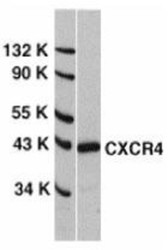
- Western Blot: CXCR4 Antibody [NBP1-77067] - HeLa whole cell lysate with CXCR4 antibody at 0.5 ug/ml.

- Submitted by
- Novus Biologicals (provider)
- Main image

- Experimental details
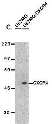
- Western Blot: CXCR4 Antibody [NBP1-77067] - U87MG and U87MG-CXCR4 extracts were included as negative and positive controls, respectively, for CXCR4 detection with anti-CXCR4 antibodies.
- Submitted by
- Novus Biologicals (provider)
- Main image
- Experimental details
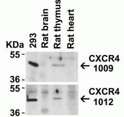
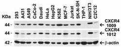
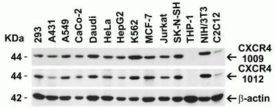
- Western Blot: CXCR4 Antibody [NBP1-77067] - Loading: Lysates/proteins at 20 ug per lane. Antibodies: 1009 (2 ug/mL) or 1012 (2 ug/mL). 1 h incubation at RT in 5% NFDM/TBST. Secondary: Goat anti-rabbit IgG HRP conjugate at 1:10000 dilution.
- Submitted by
- Novus Biologicals (provider)
- Main image
- Experimental details
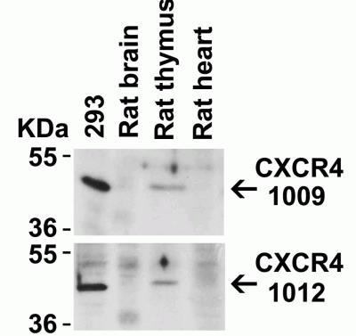
- Western Blot: CXCR4 Antibody [NBP1-77067] - Loading: 15 ug of lysates per lane. Antibodies: 1009 (1 ug/mL), 1012 (1 ug/mL), and beta-actin (1 ug/mL), 1 h incubation at RT in 5% NFDM/TBST. Secondary: Goat antirabbit IgG HRP
- Submitted by
- Novus Biologicals (provider)
- Main image
- Experimental details
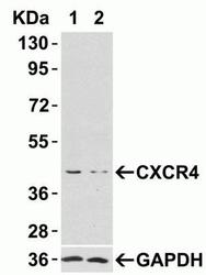
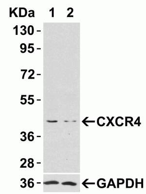
- Western Blot: CXCR4 Antibody [NBP1-77067] - HeLa cells were transfected with control siRNAs (lane 1) or CXCR4 siRNAs (lane 2) Loading: 10 ug of HeLa whole cell lysates per lane. Antibodies: 1009 (2 ug/mL), 1 h incubation at RT in 5% NFDM/TBST. Secondary: Goat antirabbit IgG HRP conjugate at 1:10000 dilution.
- Submitted by
- Novus Biologicals (provider)
- Main image
- Experimental details
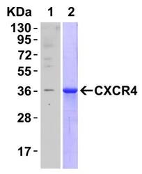
- Western Blot: CXCR4 Antibody [NBP1-77067] - Recombinant Protein Test: Loading: CXCR4 partial recombinant protein (H00007852-Q01). Lane 1: Anti-CXCR4 antibody (0.1 ug/mL) 1 h incubation at RT in 5% NFDM/TBST. Lane 2: Coomassie blue staining.

- Submitted by
- Novus Biologicals (provider)
- Main image
- Experimental details
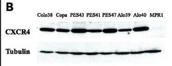
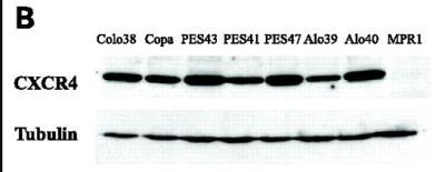
- Western Blot: CXCR4 Antibody [NBP1-77067] - CXCR4 protein was detected in the human metastatic melanoma cell lines and human melanoma cell line (colo38), but not in the human primary melanocytes (MPR1) with anti-CXCR4 antibodies.
Supportive validation
- Submitted by
- Novus Biologicals (provider)
- Main image
- Experimental details
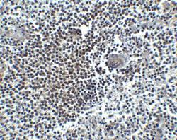
- Immunohistochemistry-Paraffin: CXCR4 Antibody [NBP1-77067] - Human spleen tissue with CXCR4 antibody at 5 ug/mL.

Supportive validation
- Submitted by
- Novus Biologicals (provider)
- Main image
- Experimental details
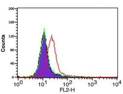
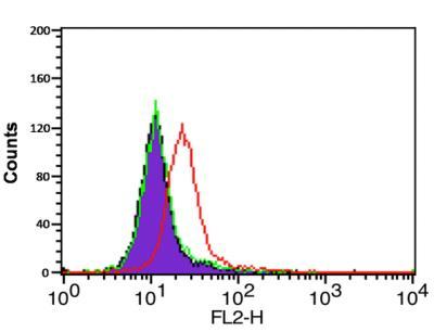
- Flow Cytometry: CXCR4 Antibody [NBP1-77067] - Analysis of HeLa cells using CXCR4 antibody at 0.1 ug/ml. Purple: cells without staining, Green: Isotype control. Red : CXCR4 antibody.
- Submitted by
- Novus Biologicals (provider)
- Main image
- Experimental details
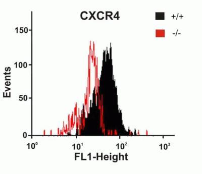
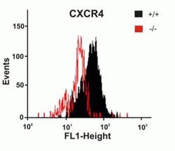
- Flow Cytometry: CXCR4 Antibody [NBP1-77067] - Astrocytes from wild-type or CXCR4 knockout mice were stained with primary antibodies against CXCR4 and FITClabeled secondary antibodies, and subsequently subjected to flow cytometry. CXCR4-/- astrocytes (red) showed loss of CXCR4 cell-surface expression compared with wild-type cells (black).