Explore
 Validate
 Learn
Learn Western blot
 ELISA
ELISA Immunocytochemistry
Antibody data
- Antibody Data
- Antigen structure
- References [4]
- Comments [0]
- Validations
- Immunocytochemistry [6]
- Immunohistochemistry [1]
Submit
Validation data
Reference
Comment
Report error
- Product number
- MA1-754 - Provider product page

- Provider
- Invitrogen Antibodies
- Product name
- PSEN2 Monoclonal Antibody (APS 26)
- Antibody type
- Monoclonal
- Antigen
- Synthetic peptide
- Description
- MA1-754 detects presenilin 2 protein (PS2) from rat, mouse, human, and nonhuman primate samples. No cross-reactivity is seen with presenilin 1. MA1-754 has successfully been used in immuno-fluorescence, immunocytochemistry, Western blot and ELISA procedures. By Western blot, this antibody detects an ~20 kDa protein representing PS2 CT (C-terminal fragment) and the ~45 kDa full-length PS2 from transfected COS-7 cells. In 4% paraformaldehyde fixed RAW cells, MA1-754 showed specific Golgi and ER labeling. The MA1-754 immunogen is a synthetic peptide corresponding to residues C L(317) P Y D P E M E E D S Y D S F G E P(334) of human PS2.
- Reactivity
- Human, Mouse, Rat
- Host
- Mouse
- Isotype
- IgG
- Antibody clone number
- APS 26
- Vial size
- 200 μg
- Concentration
- 1 mg/mL
- Storage
- -20°C, Avoid Freeze/Thaw Cycles
Submitted references Analysis of presenilin 1 and presenilin 2 expression and processing by newly developed monoclonal antibodies.
Analysis of presenilin 1 and presenilin 2 expression and processing by newly developed monoclonal antibodies.
Abrogation of the presenilin 1/beta-catenin interaction and preservation of the heterodimeric presenilin 1 complex following caspase activation.
Abrogation of the presenilin 1/beta-catenin interaction and preservation of the heterodimeric presenilin 1 complex following caspase activation.
Diehlmann A, Ida N, Weggen S, Grünberg J, Haass C, Masters CL, Bayer TA, Beyreuther K
Journal of neuroscience research 1999 May 15;56(4):405-19
Journal of neuroscience research 1999 May 15;56(4):405-19
Analysis of presenilin 1 and presenilin 2 expression and processing by newly developed monoclonal antibodies.
Diehlmann A, Ida N, Weggen S, Grünberg J, Haass C, Masters CL, Bayer TA, Beyreuther K
Journal of neuroscience research 1999 May 15;56(4):405-19
Journal of neuroscience research 1999 May 15;56(4):405-19
Abrogation of the presenilin 1/beta-catenin interaction and preservation of the heterodimeric presenilin 1 complex following caspase activation.
Tesco G, Kim TW, Diehlmann A, Beyreuther K, Tanzi RE
The Journal of biological chemistry 1998 Dec 18;273(51):33909-14
The Journal of biological chemistry 1998 Dec 18;273(51):33909-14
No comments: Submit comment
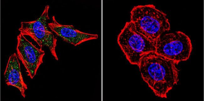
Supportive validation
- Submitted by
- Invitrogen Antibodies (provider)
- Main image

- Experimental details
- Immunofluorescent analysis of Presenilin 2 using Anti-Presenilin 2 Monoclonal Antibody (APS 26) (Product # MA1-754) shows staining in A2058 Cells. Presenilin 2 staining (green), F-Actin staining with Phalloidin (red) and nuclei with DAPI (blue) is shown. Cells were grown on chamber slides and fixed with formaldehyde prior to staining. Cells were probed without (control) or with or an antibody recognizing Presenilin 2 (Product # MA1-754) at a dilution of 1:20 over night at 4°C, washed with PBS and incubated with a DyLight-488 conjugated secondary antibody (Product # 35503, Goat Anti-Mouse). Images were taken at 60X magnification.

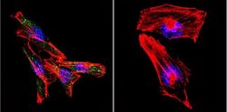
- Submitted by
- Invitrogen Antibodies (provider)
- Main image

- Experimental details
- Immunofluorescent analysis of Presenilin 2 using Anti-Presenilin 2 Monoclonal Antibody (APS 26) (Product # MA1-754) shows staining in Hela Cells. Presenilin 2 staining (green), F-Actin staining with Phalloidin (red) and nuclei with DAPI (blue) is shown. Cells were grown on chamber slides and fixed with formaldehyde prior to staining. Cells were probed without (control) or with or an antibody recognizing Presenilin 2 (Product # MA1-754) at a dilution of 1:20 over night at 4°C, washed with PBS and incubated with a DyLight-488 conjugated secondary antibody (Product # 35503, Goat Anti-Mouse). Images were taken at 60X magnification.

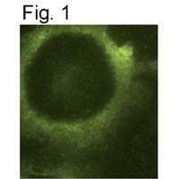
- Submitted by
- Invitrogen Antibodies (provider)
- Main image

- Experimental details
- Immunocytochemical staining of PS2 in mouse fibroblasts using Product # MA1-754.

- Submitted by
- Invitrogen Antibodies (provider)
- Main image

- Experimental details
- Immunofluorescent analysis of Presenilin 2 using Anti-Presenilin 2 Monoclonal Antibody (APS 26) (Product # MA1-754) shows staining in Hela Cells. Presenilin 2 staining (green), F-Actin staining with Phalloidin (red) and nuclei with DAPI (blue) is shown. Cells were grown on chamber slides and fixed with formaldehyde prior to staining. Cells were probed without (control) or with or an antibody recognizing Presenilin 2 (Product # MA1-754) at a dilution of 1:20 over night at 4°C, washed with PBS and incubated with a DyLight-488 conjugated secondary antibody (Product # 35503, Goat Anti-Mouse). Images were taken at 60X magnification.
- Submitted by
- Invitrogen Antibodies (provider)
- Main image
- Experimental details
- Immunofluorescent analysis of Presenilin 2 using Anti-Presenilin 2 Monoclonal Antibody (APS 26) (Product # MA1-754) shows staining in A2058 Cells. Presenilin 2 staining (green), F-Actin staining with Phalloidin (red) and nuclei with DAPI (blue) is shown. Cells were grown on chamber slides and fixed with formaldehyde prior to staining. Cells were probed without (control) or with or an antibody recognizing Presenilin 2 (Product # MA1-754) at a dilution of 1:20 over night at 4°C, washed with PBS and incubated with a DyLight-488 conjugated secondary antibody (Product # 35503, Goat Anti-Mouse). Images were taken at 60X magnification.

- Submitted by
- Invitrogen Antibodies (provider)
- Main image

- Experimental details
- Immunocytochemical staining of PS2 in mouse fibroblasts using Product # MA1-754.
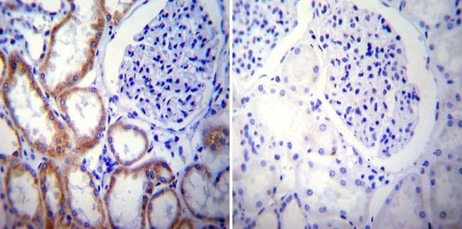
Supportive validation
- Submitted by
- Invitrogen Antibodies (provider)
- Main image
- Experimental details
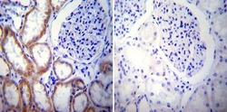
- Immunohistochemistry was performed on normal biopsies of deparaffinized Human kidney tissue. To expose target proteins, heat induced antigen retrieval was performed using 10mM sodium citrate (pH6.0) buffer, microwaved for 8-15 minutes. Following antigen retrieval tissues were blocked in 3% BSA-PBS for 30 minutes at room temperature. Tissues were then probed at a dilution of 1:20 with a mouse monoclonal antibody recognizing Presenilin 2 (Product # MA1-754) or without primary antibody (negative control) overnight at 4°C in a humidified chamber. Tissues were washed extensively with PBST and endogenous peroxidase activity was quenched with a peroxidase suppressor. Detection was performed using a biotin-conjugated secondary antibody and SA-HRP, followed by colorimetric detection using DAB. Tissues were counterstained with hematoxylin and prepped for mounting.