Antibody data
- Antibody Data
- Antigen structure
- References [1]
- Comments [0]
- Validations
- Immunohistochemistry [1]
- Other assay [1]
Submit
Validation data
Reference
Comment
Report error
- Product number
- PA5-46830 - Provider product page

- Provider
- Invitrogen Antibodies
- Product name
- GABRP Polyclonal Antibody
- Antibody type
- Polyclonal
- Antigen
- Synthetic peptide
- Description
- Peptide sequence: VEVGRSDKLS LPGFENLTAG YNKFLRPNFG GEPVQIALTL DIASISSISE Sequence homology: Dog: 100%; Horse: 100%; Human: 100%; Mouse: 100%; Rabbit: 100%; Rat: 100%
- Reactivity
- Human
- Host
- Rabbit
- Isotype
- IgG
- Vial size
- 100 μL
- Concentration
- 0.5 mg/mL
- Storage
- -20°C, Avoid Freeze/Thaw Cycles
Submitted references GABRP is a potential prognostic biomarker and correlated with immune infiltration and tumor microenvironment in pancreatic cancer.
Yang Y, Ren L, Li S, Zheng X, Liu J, Li W, Fu W, Wang J, Du G
Translational cancer research 2022 Apr;11(4):649-668
Translational cancer research 2022 Apr;11(4):649-668
No comments: Submit comment
Supportive validation
- Submitted by
- Invitrogen Antibodies (provider)
- Main image

- Experimental details
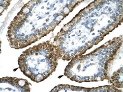
- Immunohistochemistry analysis of human intestine tissue using an anti-GABRP polyclonal antibody (Product # PA5-46830).
Supportive validation
- Submitted by
- Invitrogen Antibodies (provider)
- Main image

- Experimental details
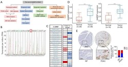
- Figure 1 GABRP mRNA expression was upregulated in pancreatic cancer. (A) The flowchart of study procedures; (B) the expression level of GABRP was measured in different types of tumor tissues and compared with normal tissues in the GEPIA2 database; (C) the expression level of GABRP was measured in different types of tumor tissues and normal tissues using the Oncomine database (P value is 0.001, fold change is 2, and gene rankings apply to all); (D) GABRP expression in tumors and matching normal tissues using two independent cohorts (GSE15471, n=78; GSE16515, n=52) derived from Gene Expression Omnibus datasets; (E) immunohistochemical analysis of GABRP expression in a human PDAC tissue microarray. Representative GABRP images are shown in the left panel. The percentages of tissues displaying low and high staining in normal pancreas and PDAC tissues are shown in the middle panel. ****, P
 Explore
 Validate
 Learn
Learn Western blot
 Immunohistochemistry