Explore
 Validate
 Learn
Learn Western blot
 Immunocytochemistry
Antibody data
- Antibody Data
- Antigen structure
- References [6]
- Comments [0]
- Validations
- Immunocytochemistry [1]
Submit
Validation data
Reference
Comment
Report error
- Product number
- HPA027407 - Provider product page

- Provider
- Atlas Antibodies
- Proper citation
- Atlas Antibodies Cat#HPA027407, RRID:AB_10959659
- Product name
- Anti-CHCHD2
- Antibody type
- Polyclonal
- Description
- Polyclonal Antibody against Human CHCHD2, Gene description: coiled-coil-helix-coiled-coil-helix domain containing 2, Alternative Gene Names: C7orf17, Validated applications: IHC, ICC, WB, Uniprot ID: Q9Y6H1, Storage: Store at +4°C for short term storage. Long time storage is recommended at -20°C.
- Reactivity
- Human
- Host
- Rabbit
- Conjugate
- Unconjugated
- Isotype
- IgG
- Vial size
- 100 µl
- Concentration
- 0.2 mg/ml
- Storage
- Store at +4°C for short term storage. Long time storage is recommended at -20°C.
- Handling
- The antibody solution should be gently mixed before use.
Submitted references Increased CHCHD2 expression promotes liver fibrosis in nonalcoholic steatohepatitis via Notch/osteopontin signaling
Loss of CHCHD2 and CHCHD10 activates OMA1 peptidase to disrupt mitochondrial cristae phenocopying patient mutations
PD-linked CHCHD2 mutations impair CHCHD10 and MICOS complex leading to mitochondria dysfunction
Loss of CHCHD10–CHCHD2 complexes required for respiration underlies the pathogenicity of a CHCHD10 mutation in ALS
CHCHD2 accumulates in distressed mitochondria and facilitates oligomerization of CHCHD10
The mitochondrial protein CHCHD2 primes the differentiation potential of human induced pluripotent stem cells to neuroectodermal lineages
Li Y, Xiu W, Xu J, Chen X, Wang G, Duan J, Sun L, Liu B, Xie W, Pu G, Wang Q, Wang C
JCI Insight 2022;7(23)
JCI Insight 2022;7(23)
Loss of CHCHD2 and CHCHD10 activates OMA1 peptidase to disrupt mitochondrial cristae phenocopying patient mutations
Narendra D, Sekine S, Poulton J, Springer D, Dombi E, Wu B, Shammas M, Nguyen D, Huang X, Liu Y
Human Molecular Genetics 2020;29(9):1547-1567
Human Molecular Genetics 2020;29(9):1547-1567
PD-linked CHCHD2 mutations impair CHCHD10 and MICOS complex leading to mitochondria dysfunction
Zhou W, Ma D, Sun A, Tran H, Ma D, Singh B, Zhou J, Zhang J, Wang D, Zhao Y, Yen P, Goh E, Tan E
Human Molecular Genetics 2019;28(7):1100-1116
Human Molecular Genetics 2019;28(7):1100-1116
Loss of CHCHD10–CHCHD2 complexes required for respiration underlies the pathogenicity of a CHCHD10 mutation in ALS
Straub I, Janer A, Weraarpachai W, Zinman L, Robertson J, Rogaeva E, Shoubridge E
Human Molecular Genetics 2018;27(1):178-189
Human Molecular Genetics 2018;27(1):178-189
CHCHD2 accumulates in distressed mitochondria and facilitates oligomerization of CHCHD10
Huang X, Wu B, Nguyen D, Liu Y, Marani M, Hench J, Bénit P, Kozjak-Pavlovic V, Rustin P, Frank S, Narendra D
Human Molecular Genetics 2018
Human Molecular Genetics 2018
The mitochondrial protein CHCHD2 primes the differentiation potential of human induced pluripotent stem cells to neuroectodermal lineages
Zhu L, Gomez-Duran A, Saretzki G, Jin S, Tilgner K, Melguizo-Sanchis D, Anyfantis G, Al-Aama J, Vallier L, Chinnery P, Lako M, Armstrong L
Journal of Cell Biology 2016;215(2):187-202
Journal of Cell Biology 2016;215(2):187-202
No comments: Submit comment
Supportive validation
- Submitted by
- Atlas Antibodies (provider)
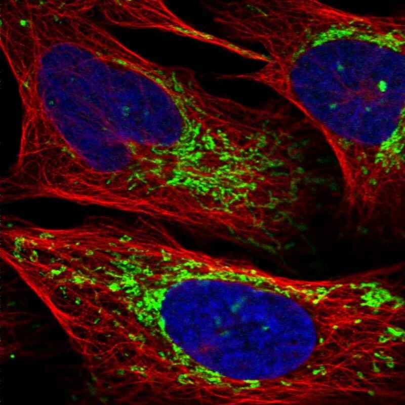
- Main image

- Experimental details
- Immunofluorescent staining of human cell line U-2 OS shows localization to mitochondria.
- Sample type
- Human