Explore
 Validate
 Learn
Learn Western blot
Antibody data
- Antibody Data
- Antigen structure
- References [0]
- Comments [0]
- Validations
- Western blot [2]
- Immunocytochemistry [1]
- Immunohistochemistry [8]
- Flow cytometry [2]
Submit
Validation data
Reference
Comment
Report error
- Product number
- GTX84242 - Provider product page

- Provider
- GeneTex
- Proper citation
- GeneTex Cat#GTX84242, RRID:AB_10729665
- Product name
- L1CAM antibody [2A6]
- Antibody type
- Monoclonal
- Reactivity
- Human
- Host
- Mouse
- Storage
- For short-term storage, store at 4¢XC or aliquot into working amounts and store at -20¢XC. For long-term storage, store at -70¢XC (aliquotted). Avoid repeated freeze-thaw cycles.
No comments: Submit comment
Supportive validation
- Submitted by
- GeneTex (provider)
- Main image
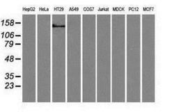
- Experimental details
- Western blot analysis of extracts (35ug) from 9 different cell lines by using anti-L1CAM monoclonal antibody.

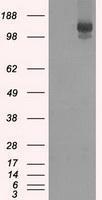
- Submitted by
- GeneTex (provider)
- Main image
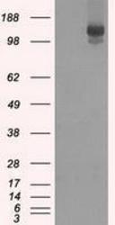
- Experimental details
- HEK293T cells were transfected with the pCMV6-ENTRY control (Left lane) or pCMV6-ENTRY L1CAM (Right lane) cDNA for 48 hrs and lysed. Equivalent amounts of cell lysates (5 ug per lane) were separated by SDS-PAGE and immunoblotted with anti-L1CAM.
Supportive validation
- Submitted by
- GeneTex (provider)
- Main image

- Experimental details
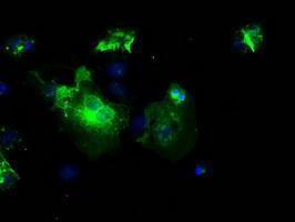
- Anti-L1CAM mouse monoclonal antibody (GTX84242) immunofluorescent staining of COS7 cells transiently transfected with L1CAM
Supportive validation
- Submitted by
- GeneTex (provider)
- Main image
- Experimental details
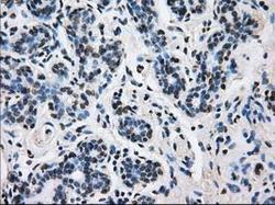
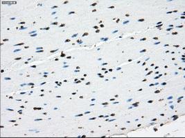
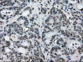
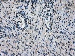
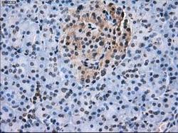
- Immunohistochemical staining of paraffin-embedded breast tissue using anti-L1CAM mouse monoclonal antibody. (GTX84242, Dilution 1:50)
- Submitted by
- GeneTex (provider)
- Main image
- Experimental details
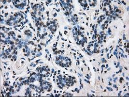
- Immunohistochemical staining of paraffin-embedded colon tissue using anti-L1CAMmouse monoclonal antibody. (GTX84242, Dilution 1:50)
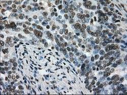
- Submitted by
- GeneTex (provider)
- Main image
- Experimental details
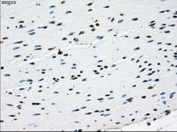
- Immunohistochemical staining of paraffin-embedded endometrium tissue using anti-L1CAMmouse monoclonal antibody. (GTX84242, Dilution 1:50)
- Submitted by
- GeneTex (provider)
- Main image
- Experimental details
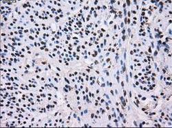
- Immunohistochemical staining of paraffin-embedded Adenocarcinoma of colon tissue using anti-L1CAMmouse monoclonal antibody. (GTX84242, Dilution 1:50)
- Submitted by
- GeneTex (provider)
- Main image
- Experimental details
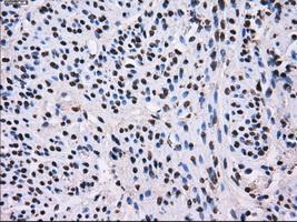
- Immunohistochemical staining of paraffin-embedded Adenocarcinoma of ovary tissue using anti-L1CAMmouse monoclonal antibody. (GTX84242, Dilution 1:50)

- Submitted by
- GeneTex (provider)
- Main image
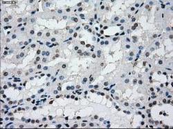
- Experimental details
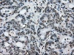
- Immunohistochemical staining of paraffin-embedded Kidney tissue using anti-L1CAMmouse monoclonal antibody. (GTX84242, Dilution 1:50)
- Submitted by
- GeneTex (provider)
- Main image
- Experimental details
- Immunohistochemical staining of paraffin-embedded Ovary tissue using anti-L1CAMmouse monoclonal antibody. (GTX84242, Dilution 1:50)

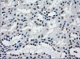
- Submitted by
- GeneTex (provider)
- Main image
- Experimental details
- Immunohistochemical staining of paraffin-embedded pancreas tissue using anti-L1CAMmouse monoclonal antibody. (GTX84242, Dilution 1:50)

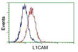
Supportive validation
- Submitted by
- GeneTex (provider)
- Main image

- Experimental details
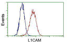
- Flow cytometric analysis of Hela cells, using anti-L1CAM antibody(GTX84242),(Red) compared to a nonspecific negative control antibody(TA50011)(Blue).
- Submitted by
- GeneTex (provider)
- Main image

- Experimental details
- Flow cytometric analysis of Jurkat cells, using anti-L1CAM antibody(GTX84242),(Red) compared to a nonspecific negative control antibody(TA50011)(Blue).