 Explore
 Validate
 Learn
Learn12-1719-42
antibody from Invitrogen Antibodies
Targeting: L1CAM
CD171, HSAS, HSAS1, MASA, MIC5, S10, SPG1
 Flow cytometry
Antibody data
- Antibody Data
- Antigen structure
- References [6]
- Comments [0]
- Validations
- Flow cytometry [2]
- Other assay [1]
Submit
Validation data
Reference
Comment
Report error
- Product number
- 12-1719-42 - Provider product page

- Provider
- Invitrogen Antibodies
- Product name
- CD171 Monoclonal Antibody (eBio5G3 (5G3)), PE, eBioscience™
- Antibody type
- Monoclonal
- Antigen
- Other
- Description
- Description: The monoclonal antibody eBio5G3 recognizes CD171 also known as neural cell adhesion molecule L1. CD171 is a member of the Ig superfamily containing 6 extracellular Ig domains and five fibronectin type III-like repeats. CD171 has been shown to function as a cell adhesion molecule mediating homotypic and heterotypic cell-cell interactions in neuronal myelination, neurite outgrowth and regeneration. Expression of CD171 has been found on monocytes and mature monocytic-derived and follicular DCs, a minor subset of lymphocytes in addition to that found on neuronal tissue and some tumor cells lines. Expression of CD171 on tumors is thought to contribute to tumor progression. Epitope of eBio5G3 is in amino-terminal Ig-like domain. Applications Reported: This eBio5G3 (5G3) antibody has been reported for use in flow cytometric analysis. Applications Tested: This eBio5G3 (5G3) antibody has been pre-titrated and tested by flow cytometric analysis of tumor cell line Panc-1. This can be used at 5 µL (0.25 µg) per test. A test is defined as the amount (µg) of antibody that will stain a cell sample in a final volume of 100 µL. Cell number should be determined empirically but can range from 10^5 to 10^8 cells/test. Excitation: 488-561 nm; Emission: 578 nm; Laser: Blue Laser, Green Laser, Yellow-Green Laser. Filtration: 0.2 µm post-manufacturing filtered.
- Reactivity
- Human
- Host
- Mouse
- Conjugate
- Yellow dye
- Isotype
- IgG
- Antibody clone number
- eBio5G3 (5G3)
- Vial size
- 100 Tests
- Concentration
- 5 µL/Test
- Storage
- 4° C, store in dark, DO NOT FREEZE!
Submitted references Evaluation of blood-based, extracellular vesicles as biomarkers for aging-related TDP-43 pathology.
Plasma Levels of Neuron- and Astrocyte-Derived Exosomal Amyloid Beta1-42, Amyloid Beta1-40, and Phosphorylated Tau Levels in Schizophrenia Patients and Non-psychiatric Comparison Subjects: Relationships With Cognitive Functioning and Psychopathology.
Human Tridimensional Neuronal Cultures for Phenotypic Drug Screening in Inherited Peripheral Neuropathies.
Human Mast Cell Proteome Reveals Unique Lineage, Putative Functions, and Structural Basis for Cell Ablation.
Comprehensive Cell Surface Antigen Analysis Identifies Transferrin Receptor Protein-1 (CD71) as a Negative Selection Marker for Human Neuronal Cells.
Knockdown of L1CAM significantly reduces metastasis in a xenograft model of human melanoma: L1CAM is a potential target for anti-melanoma therapy.
Winston CN, Sukreet S, Lynch H, Lee VM, Wilcock DM, Nelson PT, Rissman RA
Alzheimer's & dementia (Amsterdam, Netherlands) 2022;14(1):e12365
Alzheimer's & dementia (Amsterdam, Netherlands) 2022;14(1):e12365
Plasma Levels of Neuron- and Astrocyte-Derived Exosomal Amyloid Beta1-42, Amyloid Beta1-40, and Phosphorylated Tau Levels in Schizophrenia Patients and Non-psychiatric Comparison Subjects: Relationships With Cognitive Functioning and Psychopathology.
Lee EE, Winston-Gray C, Barlow JW, Rissman RA, Jeste DV
Frontiers in psychiatry 2020;11:532624
Frontiers in psychiatry 2020;11:532624
Human Tridimensional Neuronal Cultures for Phenotypic Drug Screening in Inherited Peripheral Neuropathies.
Maciel R, Correa R, Bosso Taniguchi J, Prufer Araujo I, Saporta MA
Clinical pharmacology and therapeutics 2020 May;107(5):1231-1239
Clinical pharmacology and therapeutics 2020 May;107(5):1231-1239
Human Mast Cell Proteome Reveals Unique Lineage, Putative Functions, and Structural Basis for Cell Ablation.
Plum T, Wang X, Rettel M, Krijgsveld J, Feyerabend TB, Rodewald HR
Immunity 2020 Feb 18;52(2):404-416.e5
Immunity 2020 Feb 18;52(2):404-416.e5
Comprehensive Cell Surface Antigen Analysis Identifies Transferrin Receptor Protein-1 (CD71) as a Negative Selection Marker for Human Neuronal Cells.
Menon V, Thomas R, Elgueta C, Horl M, Osborn T, Hallett PJ, Bartos M, Isacson O, Pruszak J
Stem cells (Dayton, Ohio) 2019 Oct;37(10):1293-1306
Stem cells (Dayton, Ohio) 2019 Oct;37(10):1293-1306
Knockdown of L1CAM significantly reduces metastasis in a xenograft model of human melanoma: L1CAM is a potential target for anti-melanoma therapy.
Ernst AK, Putscher A, Samatov TR, Suling A, Galatenko VV, Shkurnikov MY, Knyazev EN, Tonevitsky AG, Haalck T, Lange T, Maar H, Schröder-Schwarz J, Riecken K, Schumacher U, Wicklein D
PloS one 2018;13(2):e0192525
PloS one 2018;13(2):e0192525
No comments: Submit comment
Supportive validation
- Submitted by
- Invitrogen Antibodies (provider)
- Main image
- Experimental details
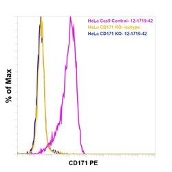
- Knockout of CD171 was achieved by CRISPR-Cas9 genome editing using LentiArray™ Lentiviral sgRNA (Product # A32042, Assay ID CRISPR651984_LV) and LentiArray Cas9 Lentivirus (Product # A32064). Flow cytometry analysis of CD171 was performed by staining HeLa CD171 Knock out cells with 0.25 µg Mouse IgG2a kappa Isotype Control (eBM2a), PE, eBioscience™ (Product # 12-4724-82, yellow histogram) or 0.25 µg CD171 Monoclonal Antibody (eBio5G3 (5G3)), PE, eBioscience™ (Product # 12-1719-42, blue histogram). HeLa Cas9 control cells were also stained with0.25 µg CD171 Monoclonal Antibody (eBio5G3 (5G3)), PE, eBioscience™ (Product # 12-1719-42, pink histogram). Lossof signal was observed in the CD171 KOcells stained with CD171 antibody clone eBio5G3 (5G3) but not in the control Cas9cells. Viable cells were used for analysis, as determined by Fixable Viability DyeeFluor™780 (Product # 65-0865-18).
- Conjugate
- Yellow dye

- Submitted by
- Invitrogen Antibodies (provider)
- Main image
- Experimental details
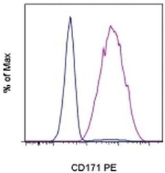
- Staining of the Panc-1 cell line with Mouse IgG2a K Isotype Control PE (Product # 12-4724-81) (blue histogram) or Anti-Human CD171 PE (purple histogram). Total viable cells were used for analysis.
- Conjugate
- Yellow dye

Supportive validation
- Submitted by
- Invitrogen Antibodies (provider)
- Main image

- Experimental details
- Figure 1 Characterization of neuronal (NDE) and astrocyte derived exosomes (ADE)s. (A) FACS analysis of plasma NDEs and ADEs following the formation of bead-antibody-exosome (BAE)-FITC complexes. Streptavidin magnetic beads were incubated with exosomes isolated from non-psychiatric comparison subjects (NC) and people with schizophrenia (PWS) ( n = 60) and enriched against biotinylated anti-human CD171 biotin (L1CAM, NDE) or anti-GLAST antibody (ADE). BAE complexes are stained with FITC prior to FACS. (B) Plasma NDE and ADE preparations from NC and PWS patients were probed with exosome marker, Flotilin-1 (1:1000). Non-exosome fraction (supernant resulting from 1 h spin at 1500G) served as the negative control while total exosomes (diluted in 1x PBS prior to neuronal and astrocyte enrichment) served as the positive control. The resultant Western Blot demonstrated that NDEs are Flotillin and Neun positive and GFAP negative. However, we were not able to get a signal for the ADEs. Due to multiple freeze-thaw cycles, the integrity of the samples used in the current study had diminished significantly.
- Conjugate
- Yellow dye
