 Explore
 Validate
 Learn
Learn13-1719-80
antibody from Invitrogen Antibodies
Targeting: L1CAM
CD171, HSAS, HSAS1, MASA, MIC5, S10, SPG1
 Western blot
 Flow cytometry
Antibody data
- Antibody Data
- Antigen structure
- References [21]
- Comments [0]
- Validations
- Flow cytometry [1]
- Other assay [6]
Submit
Validation data
Reference
Comment
Report error
- Product number
- 13-1719-80 - Provider product page

- Provider
- Invitrogen Antibodies
- Product name
- CD171 Monoclonal Antibody (eBio5G3 (5G3)), Biotin, eBioscience™
- Antibody type
- Monoclonal
- Antigen
- Other
- Description
- Description: The monoclonal antibody eBio5G3 recognizes CD171 also known as neural cell adhesion molecule L1. CD171 is a member of the Ig superfamily containing 6 extracellular Ig domains and five fibronectin type III-like repeats. CD171 has been shown to function as a cell adhesion molecule mediating homotypic and heterotypic cell-cell interactions in neuronal myelination, neurite outgrowth and regeneration. Expression of CD171 has been found on monocytes and mature monocytic-derived and follicular DCs, a minor subset of lymphocytes in addition to that found on neuronal tissue and some tumor cells lines. Expression of CD171 on tumors is thought to contribute to tumor progression. Epitope of eBio5G3 is in amino-terminal Ig-like domain. Applications Reported: This eBio5G3 (5G3) antibody has been reported for use in flow cytometric analysis, immunoblotting (WB), and immunohistology staining of frozen tissue sections. Applications Tested: This eBio5G3 (5G3) antibody has been tested by flow cytometric analysis of Panc-1 cell line. This can be used at less than or equal to 0.5 µg per test. A test is defined as the amount (µg) of antibody that will stain a cell sample in a final volume of 100 µL. Cell number should be determined empirically but can range from 10^5 to 10^8 cells/test. It is recommended that the antibody be carefully titrated for optimal performance in the assay of interest. Filtration: 0.2 µm post-manufacturing filtered.
- Reactivity
- Human
- Host
- Mouse
- Conjugate
- Biotin
- Isotype
- IgG
- Antibody clone number
- eBio5G3 (5G3)
- Vial size
- 25 µg
- Concentration
- 0.5 mg/mL
- Storage
- 4° C, store in dark, DO NOT FREEZE!
Submitted references L1CAM immunocapture generates a unique extracellular vesicle population with a reproducible miRNA fingerprint.
Evaluation of blood-based, extracellular vesicles as biomarkers for aging-related TDP-43 pathology.
Human neural cell type-specific extracellular vesicle proteome defines disease-related molecules associated with activated astrocytes in Alzheimer's disease brain.
Neuronally-enriched exosomal microRNA-27b mediates acute effects of ibuprofen on reward-related brain activity in healthy adults: a randomized, placebo-controlled, double-blind trial.
Insulin receptor substrate in brain-enriched exosomes in subjects with major depression: on the path of creation of biosignatures of central insulin resistance.
Characterization and Biomarker Analyses of Post-COVID-19 Complications and Neurological Manifestations.
Neuronal and Astrocytic Extracellular Vesicle Biomarkers in Blood Reflect Brain Pathology in Mouse Models of Alzheimer's Disease.
Plasma Levels of Neuron- and Astrocyte-Derived Exosomal Amyloid Beta1-42, Amyloid Beta1-40, and Phosphorylated Tau Levels in Schizophrenia Patients and Non-psychiatric Comparison Subjects: Relationships With Cognitive Functioning and Psychopathology.
An miRNA fingerprint using neural-enriched extracellular vesicles from blood plasma: towards a biomarker for amyotrophic lateral sclerosis/motor neuron disease.
Assessing Neuronal and Astrocyte Derived Exosomes From Individuals With Mild Traumatic Brain Injury for Markers of Neurodegeneration and Cytotoxic Activity.
Increased DJ-1 and α-Synuclein in Plasma Neural-Derived Exosomes as Potential Markers for Parkinson's Disease.
Detection of Aggregation-Competent Tau in Neuron-Derived Extracellular Vesicles.
Pericyte-like spreading by disseminated cancer cells activates YAP and MRTF for metastatic colonization.
Plasma Extracellular Vesicles Enriched for Neuronal Origin: A Potential Window into Brain Pathologic Processes.
Exosomal biomarkers of brain insulin resistance associated with regional atrophy in Alzheimer's disease.
Prediction of conversion from mild cognitive impairment to dementia with neuronally derived blood exosome protein profile.
Plasma neuronal exosomal levels of Alzheimer's disease biomarkers in normal aging.
Low neural exosomal levels of cellular survival factors in Alzheimer's disease.
Altered lysosomal proteins in neural-derived plasma exosomes in preclinical Alzheimer disease.
Dysfunctionally phosphorylated type 1 insulin receptor substrate in neural-derived blood exosomes of preclinical Alzheimer's disease.
Serpins promote cancer cell survival and vascular co-option in brain metastasis.
Dunlop RA, Banack SA, Cox PA
RNA biology 2023 Jan;20(1):140-148
RNA biology 2023 Jan;20(1):140-148
Evaluation of blood-based, extracellular vesicles as biomarkers for aging-related TDP-43 pathology.
Winston CN, Sukreet S, Lynch H, Lee VM, Wilcock DM, Nelson PT, Rissman RA
Alzheimer's & dementia (Amsterdam, Netherlands) 2022;14(1):e12365
Alzheimer's & dementia (Amsterdam, Netherlands) 2022;14(1):e12365
Human neural cell type-specific extracellular vesicle proteome defines disease-related molecules associated with activated astrocytes in Alzheimer's disease brain.
You Y, Muraoka S, Jedrychowski MP, Hu J, McQuade AK, Young-Pearse T, Aslebagh R, Shaffer SA, Gygi SP, Blurton-Jones M, Poon WW, Ikezu T
Journal of extracellular vesicles 2022 Jan;11(1):e12183
Journal of extracellular vesicles 2022 Jan;11(1):e12183
Neuronally-enriched exosomal microRNA-27b mediates acute effects of ibuprofen on reward-related brain activity in healthy adults: a randomized, placebo-controlled, double-blind trial.
Burrows K, Figueroa-Hall LK, Kuplicki R, Stewart JL, Alarbi AM, Ramesh R, Savitz JB, Teague TK, Risbrough VB, Paulus MP
Scientific reports 2022 Jan 17;12(1):861
Scientific reports 2022 Jan 17;12(1):861
Insulin receptor substrate in brain-enriched exosomes in subjects with major depression: on the path of creation of biosignatures of central insulin resistance.
Nasca C, Dobbin J, Bigio B, Watson K, de Angelis P, Kautz M, Cochran A, Mathé AA, Kocsis JH, Lee FS, Murrough JW, McEwen BS, Rasgon N
Molecular psychiatry 2021 Sep;26(9):5140-5149
Molecular psychiatry 2021 Sep;26(9):5140-5149
Characterization and Biomarker Analyses of Post-COVID-19 Complications and Neurological Manifestations.
Sun B, Tang N, Peluso MJ, Iyer NS, Torres L, Donatelli JL, Munter SE, Nixon CC, Rutishauser RL, Rodriguez-Barraquer I, Greenhouse B, Kelly JD, Martin JN, Deeks SG, Henrich TJ, Pulliam L
Cells 2021 Feb 13;10(2)
Cells 2021 Feb 13;10(2)
Neuronal and Astrocytic Extracellular Vesicle Biomarkers in Blood Reflect Brain Pathology in Mouse Models of Alzheimer's Disease.
Delgado-Peraza F, Nogueras-Ortiz CJ, Volpert O, Liu D, Goetzl EJ, Mattson MP, Greig NH, Eitan E, Kapogiannis D
Cells 2021 Apr 23;10(5)
Cells 2021 Apr 23;10(5)
Plasma Levels of Neuron- and Astrocyte-Derived Exosomal Amyloid Beta1-42, Amyloid Beta1-40, and Phosphorylated Tau Levels in Schizophrenia Patients and Non-psychiatric Comparison Subjects: Relationships With Cognitive Functioning and Psychopathology.
Lee EE, Winston-Gray C, Barlow JW, Rissman RA, Jeste DV
Frontiers in psychiatry 2020;11:532624
Frontiers in psychiatry 2020;11:532624
An miRNA fingerprint using neural-enriched extracellular vesicles from blood plasma: towards a biomarker for amyotrophic lateral sclerosis/motor neuron disease.
Banack SA, Dunlop RA, Cox PA
Open biology 2020 Jun;10(6):200116
Open biology 2020 Jun;10(6):200116
Assessing Neuronal and Astrocyte Derived Exosomes From Individuals With Mild Traumatic Brain Injury for Markers of Neurodegeneration and Cytotoxic Activity.
Winston CN, Romero HK, Ellisman M, Nauss S, Julovich DA, Conger T, Hall JR, Campana W, O'Bryant SE, Nievergelt CM, Baker DG, Risbrough VB, Rissman RA
Frontiers in neuroscience 2019;13:1005
Frontiers in neuroscience 2019;13:1005
Increased DJ-1 and α-Synuclein in Plasma Neural-Derived Exosomes as Potential Markers for Parkinson's Disease.
Zhao ZH, Chen ZT, Zhou RL, Zhang X, Ye QY, Wang YZ
Frontiers in aging neuroscience 2018;10:438
Frontiers in aging neuroscience 2018;10:438
Detection of Aggregation-Competent Tau in Neuron-Derived Extracellular Vesicles.
Guix FX, Corbett GT, Cha DJ, Mustapic M, Liu W, Mengel D, Chen Z, Aikawa E, Young-Pearse T, Kapogiannis D, Selkoe DJ, Walsh DM
International journal of molecular sciences 2018 Feb 27;19(3)
International journal of molecular sciences 2018 Feb 27;19(3)
Pericyte-like spreading by disseminated cancer cells activates YAP and MRTF for metastatic colonization.
Er EE, Valiente M, Ganesh K, Zou Y, Agrawal S, Hu J, Griscom B, Rosenblum M, Boire A, Brogi E, Giancotti FG, Schachner M, Malladi S, Massagué J
Nature cell biology 2018 Aug;20(8):966-978
Nature cell biology 2018 Aug;20(8):966-978
Plasma Extracellular Vesicles Enriched for Neuronal Origin: A Potential Window into Brain Pathologic Processes.
Mustapic M, Eitan E, Werner JK Jr, Berkowitz ST, Lazaropoulos MP, Tran J, Goetzl EJ, Kapogiannis D
Frontiers in neuroscience 2017;11:278
Frontiers in neuroscience 2017;11:278
Exosomal biomarkers of brain insulin resistance associated with regional atrophy in Alzheimer's disease.
Mullins RJ, Mustapic M, Goetzl EJ, Kapogiannis D
Human brain mapping 2017 Apr;38(4):1933-1940
Human brain mapping 2017 Apr;38(4):1933-1940
Prediction of conversion from mild cognitive impairment to dementia with neuronally derived blood exosome protein profile.
Winston CN, Goetzl EJ, Akers JC, Carter BS, Rockenstein EM, Galasko D, Masliah E, Rissman RA
Alzheimer's & dementia (Amsterdam, Netherlands) 2016;3:63-72
Alzheimer's & dementia (Amsterdam, Netherlands) 2016;3:63-72
Plasma neuronal exosomal levels of Alzheimer's disease biomarkers in normal aging.
Abner EL, Jicha GA, Shaw LM, Trojanowski JQ, Goetzl EJ
Annals of clinical and translational neurology 2016 May;3(5):399-403
Annals of clinical and translational neurology 2016 May;3(5):399-403
Low neural exosomal levels of cellular survival factors in Alzheimer's disease.
Goetzl EJ, Boxer A, Schwartz JB, Abner EL, Petersen RC, Miller BL, Carlson OD, Mustapic M, Kapogiannis D
Annals of clinical and translational neurology 2015 Jul;2(7):769-73
Annals of clinical and translational neurology 2015 Jul;2(7):769-73
Altered lysosomal proteins in neural-derived plasma exosomes in preclinical Alzheimer disease.
Goetzl EJ, Boxer A, Schwartz JB, Abner EL, Petersen RC, Miller BL, Kapogiannis D
Neurology 2015 Jul 7;85(1):40-7
Neurology 2015 Jul 7;85(1):40-7
Dysfunctionally phosphorylated type 1 insulin receptor substrate in neural-derived blood exosomes of preclinical Alzheimer's disease.
Kapogiannis D, Boxer A, Schwartz JB, Abner EL, Biragyn A, Masharani U, Frassetto L, Petersen RC, Miller BL, Goetzl EJ
FASEB journal : official publication of the Federation of American Societies for Experimental Biology 2015 Feb;29(2):589-96
FASEB journal : official publication of the Federation of American Societies for Experimental Biology 2015 Feb;29(2):589-96
Serpins promote cancer cell survival and vascular co-option in brain metastasis.
Valiente M, Obenauf AC, Jin X, Chen Q, Zhang XH, Lee DJ, Chaft JE, Kris MG, Huse JT, Brogi E, Massagué J
Cell 2014 Feb 27;156(5):1002-16
Cell 2014 Feb 27;156(5):1002-16
No comments: Submit comment
Supportive validation
- Submitted by
- Invitrogen Antibodies (provider)
- Main image

- Experimental details
- Staining of Panc-1 cell line with 0.25 µg of Mouse IgG2a kappa Isotype Control Biotin (Product # 13-4724-85) (blue histogram) or 0.25 µg of Anti-Human CD171 Biotin (purple histogram) followed by Streptavidin PE (Product # 12-4317-87). Total viable cells were used for analysis.
- Conjugate
- Biotin

Supportive validation
- Submitted by
- Invitrogen Antibodies (provider)
- Main image

- Experimental details
- NULL
- Conjugate
- Biotin

- Submitted by
- Invitrogen Antibodies (provider)
- Main image

- Experimental details
- NULL
- Conjugate
- Biotin

- Submitted by
- Invitrogen Antibodies (provider)
- Main image

- Experimental details
- NULL
- Conjugate
- Biotin

- Submitted by
- Invitrogen Antibodies (provider)
- Main image
- Experimental details
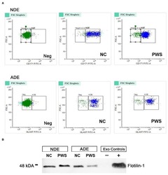
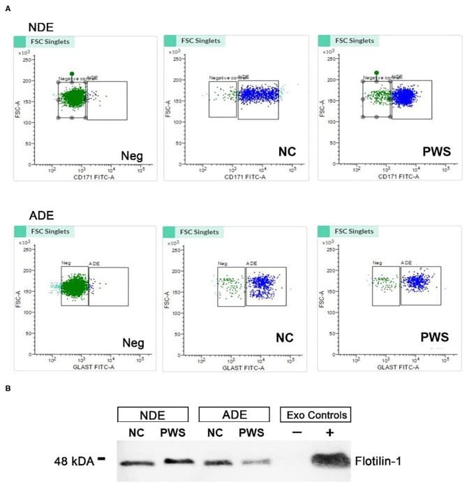
- Figure 1 Characterization of neuronal (NDE) and astrocyte derived exosomes (ADE)s. (A) FACS analysis of plasma NDEs and ADEs following the formation of bead-antibody-exosome (BAE)-FITC complexes. Streptavidin magnetic beads were incubated with exosomes isolated from non-psychiatric comparison subjects (NC) and people with schizophrenia (PWS) ( n = 60) and enriched against biotinylated anti-human CD171 biotin (L1CAM, NDE) or anti-GLAST antibody (ADE). BAE complexes are stained with FITC prior to FACS. (B) Plasma NDE and ADE preparations from NC and PWS patients were probed with exosome marker, Flotilin-1 (1:1000). Non-exosome fraction (supernant resulting from 1 h spin at 1500G) served as the negative control while total exosomes (diluted in 1x PBS prior to neuronal and astrocyte enrichment) served as the positive control. The resultant Western Blot demonstrated that NDEs are Flotillin and Neun positive and GFAP negative. However, we were not able to get a signal for the ADEs. Due to multiple freeze-thaw cycles, the integrity of the samples used in the current study had diminished significantly.
- Conjugate
- Biotin
- Submitted by
- Invitrogen Antibodies (provider)
- Main image

- Experimental details
- Figure 1. Increased number of L1CAM + exosomes in subjects with MDD as compared to controls. (A) Workflow for the isolation, enrichment and analysis of exosomes from human plasma. Magnetic beads were used for enrichment of L1CAM + (brain-enriched) exosomes based on the immunoprecipitation of the L1CAM target bound to the exosomal surface. (B) Expression array validated the accuracy of the exosome isolation and confirmed the exclusion of cellular contamination from cellular vesicles as showed by the low staining for GM130, a marker for cis-Golgi vesicles. In addition to positive (green rectangular) and negative (red rectangular) controls, the expression array detected the following known exosome markers (orange rectangular) in our set of samples: Flot-1, ICAM, Alix, CD81, CD63, EpCAM, ANXA5, TSG101. (C) Western blot analysis confirmed isolation of L1CAM + exosomes. (D) Number of L1CAM + exosomes from subjects in depressive episode during study participation (n=48) as compared to controls (n=23). * indicates significant comparisons with controls. Dashed bars indicate group mean. See also related Figure SI1 .
- Conjugate
- Biotin

- Submitted by
- Invitrogen Antibodies (provider)
- Main image

- Experimental details
- Figure 7 Neuronally-enriched exosomes (NEEs). ( A ) Schematic depiction of NEE enrichment. ( B ) Fluorescence activated cell sorting (FACS) results of NEEs and negative control (No exosomes). ( C ) Western blot analysis of NEEs, total exosomes (TEs), exosome-depleted serum, and negative controls (No exosomes) with anti-CD171 antibody marker, image cropped was from the same gel. ( D ) Size and concentration analysis of NEEs using Nanoparticle Tracking Analysis System.
- Conjugate
- Biotin
