Explore
 Validate
 Learn
Learn Western blot
 Immunohistochemistry
Antibody data
- Antibody Data
- Antigen structure
- References [2]
- Comments [0]
- Validations
- Immunohistochemistry [1]
Submit
Validation data
Reference
Comment
Report error
- Product number
- AF277 - Provider product page

- Provider
- R&D Systems
- Product name
- Human L1CAM Antibody
- Antibody type
- Polyclonal
- Description
- Immunogen affinity purified. Detects human L1CAM in direct ELISAs and Western blots. In direct ELISAs, approximately 10% cross-reactivity with recombinant human (rh) ICAM-2 and rhICAM-3 is observed and less than 1% cross-reactvity with rhCD31 and recombinant mouse VCAM-1 is observed.
- Reactivity
- Human
- Host
- Goat
- Conjugate
- Unconjugated
- Antigen sequence
CAA42508- Isotype
- IgG
- Vial size
- 100 ug
- Concentration
- LYOPH
- Storage
- Use a manual defrost freezer and avoid repeated freeze-thaw cycles. 12 months from date of receipt, -20 to -70 °C as supplied. 1 month, 2 to 8 °C under sterile conditions after reconstitution. 6 months, -20 to -70 °C under sterile conditions after reconstitution.
Submitted references Secretome and degradome profiling shows that Kallikrein-related peptidases 4, 5, 6, and 7 induce TGFβ-1 signaling in ovarian cancer cells.
The L1 cell adhesion molecule is a potential biomarker of human distal nephron injury in acute tubular necrosis.
Shahinian H, Loessner D, Biniossek ML, Kizhakkedathu JN, Clements JA, Magdolen V, Schilling O
Molecular oncology 2014 Feb;8(1):68-82
Molecular oncology 2014 Feb;8(1):68-82
The L1 cell adhesion molecule is a potential biomarker of human distal nephron injury in acute tubular necrosis.
Allory Y, Audard V, Fontanges P, Ronco P, Debiec H
Kidney international 2008 Mar;73(6):751-8
Kidney international 2008 Mar;73(6):751-8
No comments: Submit comment
Supportive validation
- Submitted by
- R&D Systems (provider)
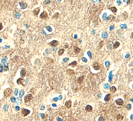
- Main image
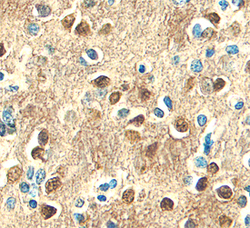
- Experimental details
- L1CAM in Human Brain. L1CAM was detected in immersion fixed paraffin-embedded sections of human brain (hippocampus) using Goat Anti-Human L1CAM Antigen Affinity-purified Poly-clonal Antibody (Catalog # AF277) at 10 µg/mL overnight at 4 °C. Before incubation with the primary antibody, tissue was subjected to heat-induced epitope retrieval using Antigen Retrieval Reagent-Basic (Catalog # CTS013). Tissue was stained using the Anti-Goat HRP-DAB Cell & Tissue Staining Kit (brown; Catalog # CTS008) and counter-stained with hematoxylin (blue). Specific staining was localized to plasma membranes and cytoplasm of neurons. View our protocol for Chromogenic IHC Staining of Paraffin-embedded Tissue Sections.