Explore
 Validate
 Learn
Learn20697-1-AP
antibody from Invitrogen Antibodies
Targeting: SHH
HHG1, HLP3, HPE3, MCOPCB5, SMMCI, TPT, TPTPS
 Western blot Immunocytochemistry
 Immunoprecipitation Immunohistochemistry Flow cytometry Other assay
Antibody data
- Antibody Data
- Antigen structure
- References [0]
- Comments [0]
- Validations
- Western blot [3]
- Immunocytochemistry [2]
- Immunohistochemistry [7]
- Flow cytometry [1]
- Other assay [1]
Submit
Validation data
Reference
Comment
Report error
- Product number
- 20697-1-AP - Provider product page

- Provider
- Invitrogen Antibodies
- Product name
- SHH Polyclonal Antibody
- Antibody type
- Polyclonal
- Antigen
- Synthetic peptide
- Reactivity
- Human, Mouse, Rat
- Host
- Rabbit
- Isotype
- IgG
- Vial size
- 150 µL
- Concentration
- 0.4 mg/mL
- Storage
- -20°C
No comments: Submit comment
Supportive validation
- Submitted by
- Invitrogen Antibodies (provider)
- Main image

- Experimental details
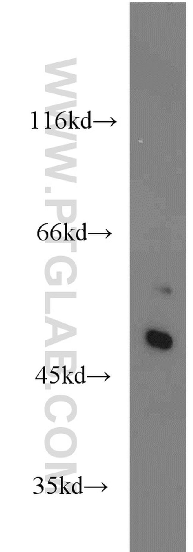
- Mouse liver tissue were subjected to SDS PAGE followed by western blot with 20697-1-AP (SHH antibody) at dilution of 1:300 incubated at room temperature for 1.5 hours.
- Submitted by
- Invitrogen Antibodies (provider)
- Main image

- Experimental details
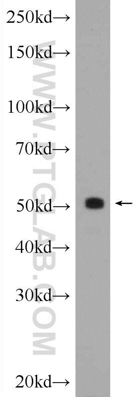
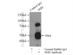
- Rat kidney tissue were subjected to SDS PAGE followed by western blot with 20697-1-AP (SHH Antibody) at dilution of 1:600 incubated at room temperature for 1.5 hours.
- Submitted by
- Invitrogen Antibodies (provider)
- Main image

- Experimental details
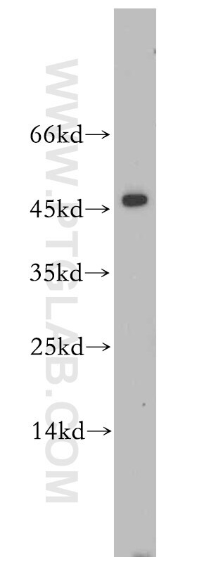
- Mouse liver tissue were subjected to SDS PAGE followed by western blot with 20697-1-AP (SHH antibody) at dilution of 1:800 incubated at room temperature for 1.5 hours.
Supportive validation
- Submitted by
- Invitrogen Antibodies (provider)
- Main image
- Experimental details
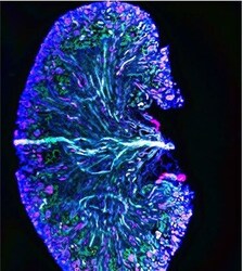
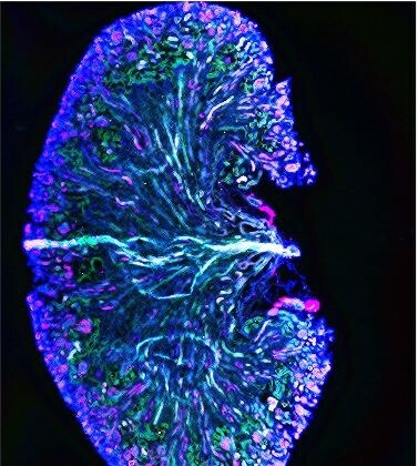
- Frozen tissue section of adult mouse kidney was stained for acetylated ɑ-tubulin (magenta, Cat. No CL488-66200), CD31/PECAM-1 (white), and Shh (green, Cat. No Product # 20697-1-AP) with DAPI as a counterstain for visualizing the nucleus (blue). acetylated ɑ-tubulin stains primary cilia and was conjugated to Coralite-488 fluorescent dye and psedocolored to magenta. CD31 stains endocardial/endothelial cells and was visualized with an Alexa Fluor 647 secondary antibody and psedocolored to white. Shh stains Sonic Hedgehog and was visualized with an Alexa Fluor 555 secondary antibody and psedocolored to green. The Image and figure legends are intellectual property of @LAF_in_the_LAB.
- Submitted by
- Invitrogen Antibodies (provider)
- Main image
- Experimental details
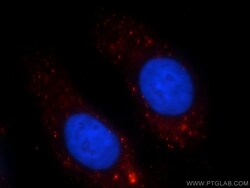
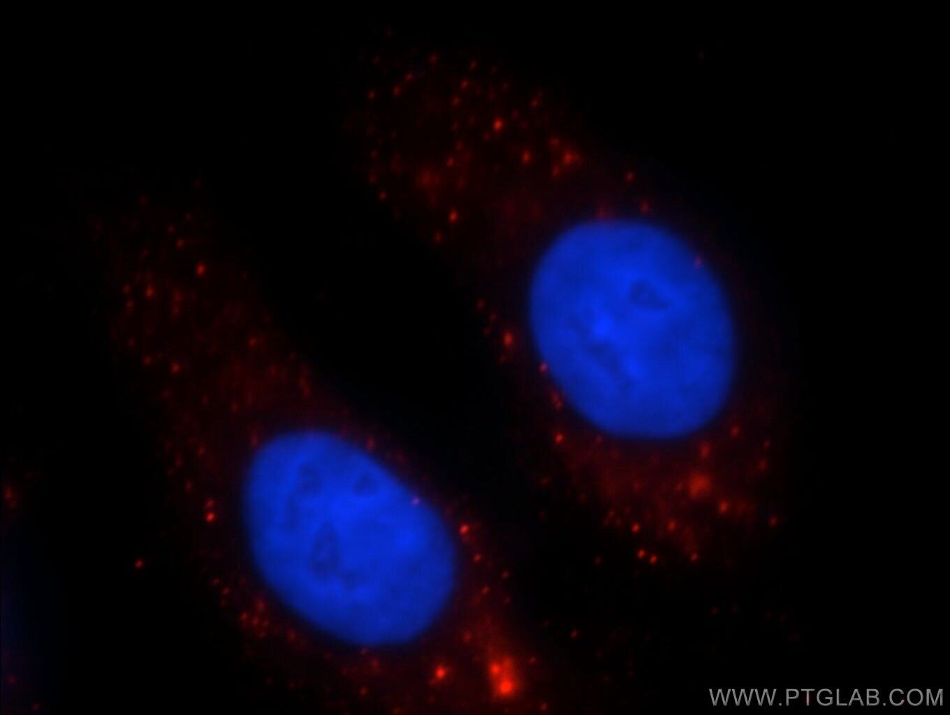
- Immunofluorescent analysis of HepG2 cells, using SHH antibody 20697-1-AP at 1:25 dilution and Rhodamine-labeled goat anti-rabbit IGG (red). Blue pseudocolor = DAPI (fluorescent DNA dye).
Supportive validation
- Submitted by
- Invitrogen Antibodies (provider)
- Main image
- Experimental details
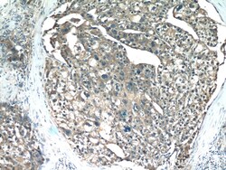
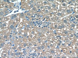
- Immunohistochemistry of paraffin-embedded human liver cancer tissue slide using 20697-1-AP (SHH Antibody) at dilution of 1:50 (under 10x lens).

- Submitted by
- Invitrogen Antibodies (provider)
- Main image

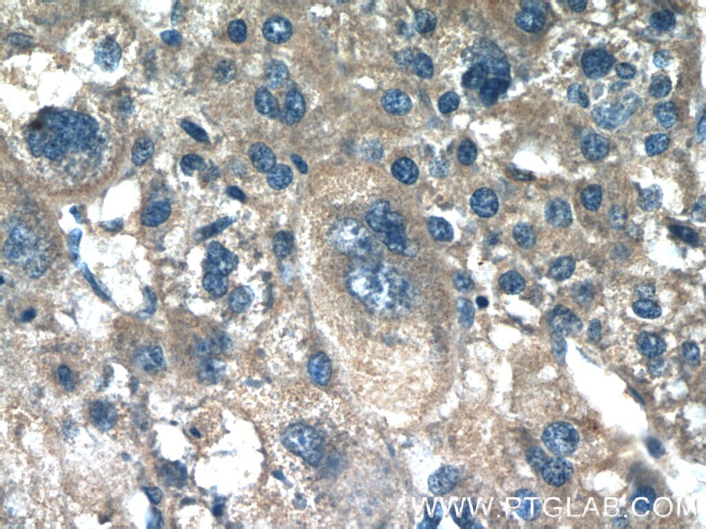
- Experimental details
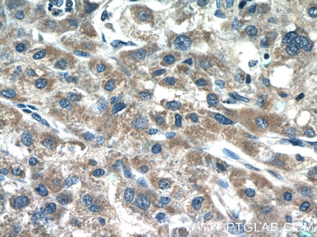
- Immunohistochemistry of paraffin-embedded human liver cancer tissue slide using 20697-1-AP (SHH Antibody) at dilution of 1:50 (under 40x lens).
- Submitted by
- Invitrogen Antibodies (provider)
- Main image
- Experimental details
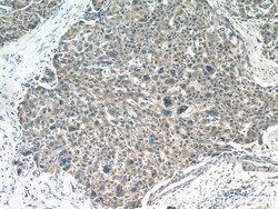
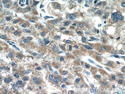
- Immunohistochemistry of paraffin-embedded human liver cancer tissue slide using 20697-1-AP (SHH Antibody) at dilution of 1:50 (under 10x lens).
- Submitted by
- Invitrogen Antibodies (provider)
- Main image
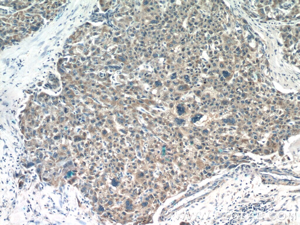
- Experimental details
- Immunohistochemistry of paraffin-embedded human liver cancer tissue slide using 20697-1-AP (SHH Antibody) at dilution of 1:50 (under 40x lens).
- Submitted by
- Invitrogen Antibodies (provider)
- Main image
- Experimental details
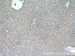
- Immunohistochemistry of paraffin-embedded mouse liver tissue slide using 20697-1-AP (SHH Antibody) at dilution of 1:50 (under 10x lens).
- Submitted by
- Invitrogen Antibodies (provider)
- Main image
- Experimental details
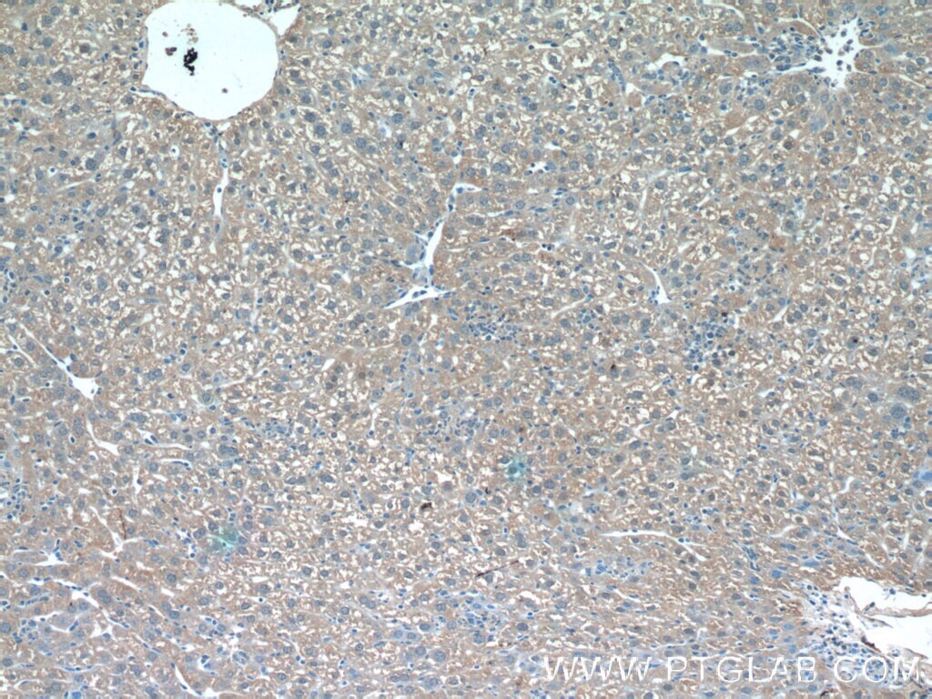
- Immunohistochemistry of paraffin-embedded mouse liver tissue slide using 20697-1-AP (SHH Antibody) at dilution of 1:50 (under 40x lens).

- Submitted by
- Invitrogen Antibodies (provider)
- Main image

- Experimental details
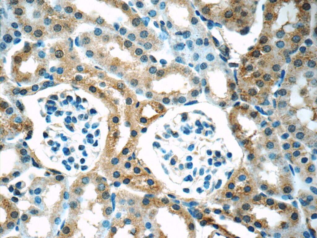
- Immunohistochemistry of paraffin-embedded mouse kidney tissue slide using 20697-1-AP ( SHH Antibody) at dilution of 1:50 (under 40x lens).
Supportive validation
- Submitted by
- Invitrogen Antibodies (provider)
- Main image
- Experimental details
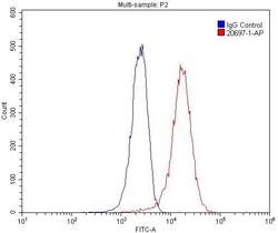
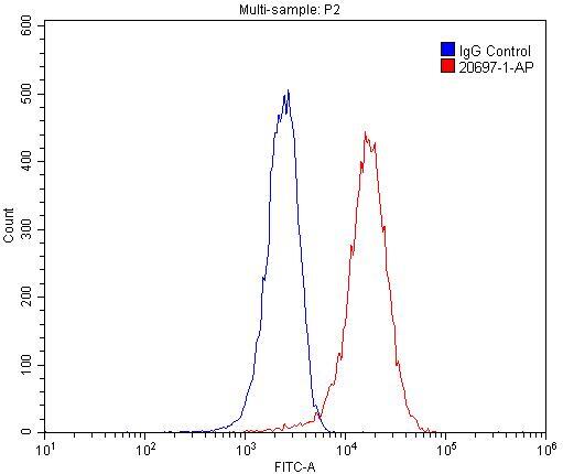
- 1X10^6 HepG2 cells were stained with 0.2ug SHH antibody (20697-1-AP, red) and control antibody (blue). Fixed with 4% PFA blocked with 3% BSA (30 min). Alexa Fluor 488-conjugated AffiniPure Goat Anti-Rabbit IGG (H+L) with dilution 1:1500.
Supportive validation
- Submitted by
- Invitrogen Antibodies (provider)
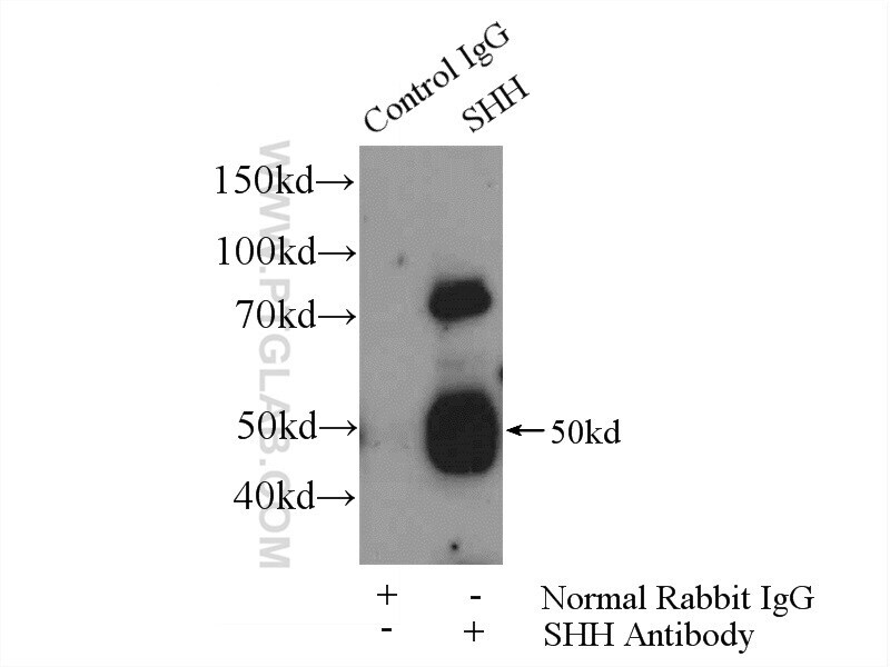
- Main image
- Experimental details
- IP result of anti-SHH (IP:20697-1-AP, 4ug; Detection:20697-1-AP 1:500) with mouse liver tissue lysate 6400ug.