Explore
 Validate
 Learn
Learn Western blot
Antibody data
- Antibody Data
- Antigen structure
- References [1]
- Comments [0]
- Validations
- Western blot [2]
- Immunocytochemistry [5]
- Immunohistochemistry [4]
- Flow cytometry [1]
- Other assay [1]
Submit
Validation data
Reference
Comment
Report error
- Product number
- MA5-32489 - Provider product page

- Provider
- Invitrogen Antibodies
- Product name
- Fas Recombinant Rabbit Monoclonal Antibody (JJ0942)
- Antibody type
- Monoclonal
- Antigen
- Recombinant full-length protein
- Description
- Recombinant rabbit monoclonal antibodies are produced using in vitro expression systems. The expression systems are developed by cloning in the specific antibody DNA sequences from immunoreactive rabbits. Then, individual clones are screened to select the best candidates for production. The advantages of using recombinant rabbit monoclonal antibodies include: better specificity and sensitivity, lot-to-lot consistency, animal origin-free formulations, and broader immunoreactivity to diverse targets due to larger rabbit immune repertoire.
- Reactivity
- Human
- Host
- Rabbit
- Isotype
- IgG
- Antibody clone number
- JJ0942
- Vial size
- 100 μL
- Concentration
- 1 mg/mL
- Storage
- Store at 4°C short term. For long term storage, store at -20°C, avoiding freeze/thaw cycles.
Submitted references Fas-Fas Ligand Interplay in the Periphery of Salivary Gland Carcinomas as a New Checkpoint Predictor for Disease Severity and Immunotherapy Response.
Strizova Z, Kuchar M, Capkova L, Komarc M, Skrivan J, Bartunkova J, Plzak J, Smrz D
Biomedicines 2021 Apr 8;9(4)
Biomedicines 2021 Apr 8;9(4)
No comments: Submit comment
Supportive validation
- Submitted by
- Invitrogen Antibodies (provider)
- Main image
- Experimental details
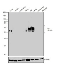
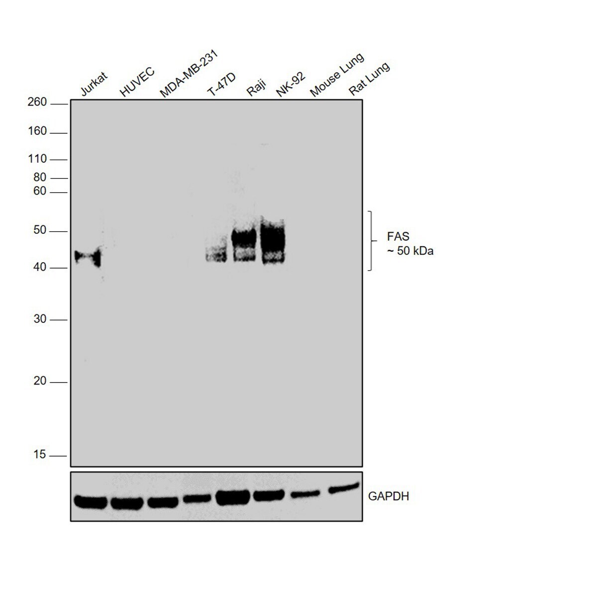
- Western Blot was performed using Anti-Fas Recombinant Rabbit Monoclonal Antibody (JJ0942) (Product # MA5-32489) and a 50 kDa band corresponding to glycosylated FAS (Tumor necrosis factor receptor superfamily member 6) was observed across cell lines tested except in HUVEC and MDA-MB-231 which are reported low expressing for the same. Whole cell extracts (40 µg lysate) of Jurkat (Lane 1), HUVEC (Lane 2), MDA-MB-231 (Lane 3), T-47D (Lane 4), Raji (Lane 5), NK-92 (Lane 6) and tissue extracts (40 µg lysate) of Mouse Lung (Lane 7) and Rat Lung (Lane 8) were electrophoresed using NuPAGE™ 10% Bis-Tris Protein Gel (Product # NP0301BOX). Resolved proteins were then transferred onto a nitrocellulose membrane (Product # IB23001) by iBlot® 2 Dry Blotting System (Product # IB21001). The blot was probed with the primary antibody (1:1000 dilution) and detected by chemiluminescence with Goat anti-Rabbit IgG (Heavy Chain) Superclonal™ Recombinant Secondary Antibody, HRP (Product # A27036, 1:10000 dilution) using the iBright FL 1000 (Product # A32752). Chemiluminescent detection was performed using SuperSignal™ West Dura Extended Duration Substrate (Product # 34076).
- Submitted by
- Invitrogen Antibodies (provider)
- Main image
- Experimental details
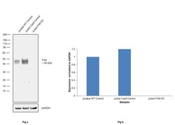
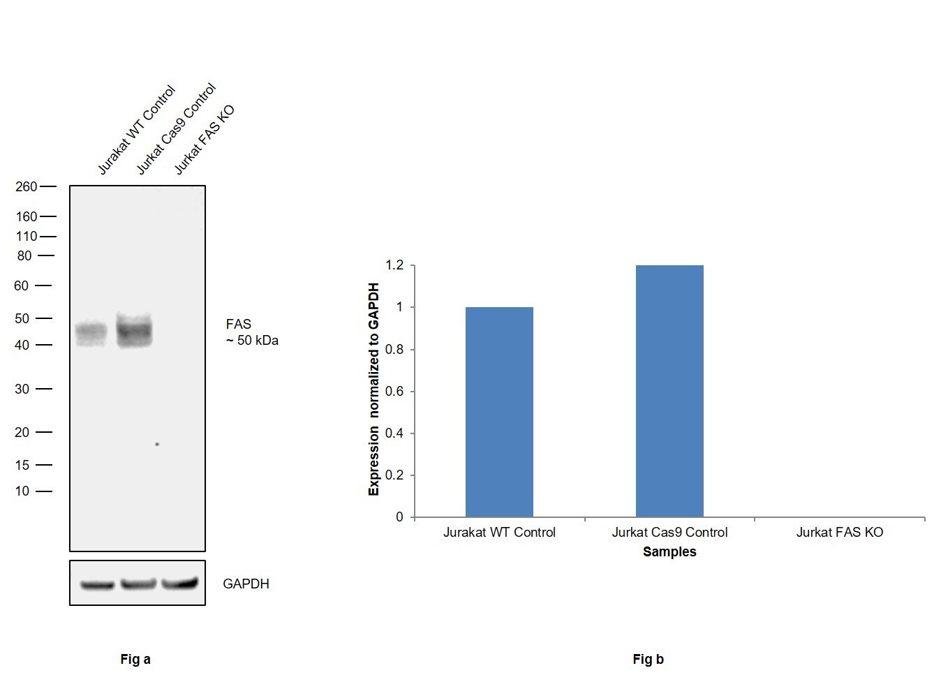
- Knockout of Fas was achieved by CRISPR-Cas9 genome editing using LentiArray™ Lentiviral sgRNA (Product # A32042, Assay ID CRISPR680010_LV) and LentiArray Cas9 Lentivirus (Product # A32064). Western blot analysis of Fas was performed by loading 30 µg of Jurkat Wild Type (Lane 1), Jurkat Cas9 (Lane 2) andJurkat FAS KO (Lane 3) membrane enriched extracts. The samples were electrophoresed using NuPAGE™ Novex™ 4-12% Bis-Tris Protein Gel (Product # NP0322BOX). Resolved proteins were then transferred onto a nitrocellulose membrane (Product # IB23001) by iBlot® 2 Dry Blotting System (Product # IB21001). The blot was probed with Anti-Fas Recombinant Rabbit Monoclonal Antibody (JJ0942) (Product # MA5-32489, 1:1,000 dilution) and Goat anti-Rabbit IgG (Heavy Chain) Superclonal™ Recombinant Secondary Antibody, HRP (Product # A27036, 1:10,000 dilution) using the iBright FL 1000 (Product # A32752). Chemiluminescent detection was performed using SuperSignal™ West Dura Extended Duration Substrate (Product # 34076). Loss of signal upon CRISPR mediated knockout (KO) using the LentiArray™ CRISPR product line confirms that antibody is specific to Fas.
Supportive validation
- Submitted by
- Invitrogen Antibodies (provider)
- Main image
- Experimental details
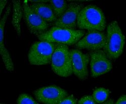
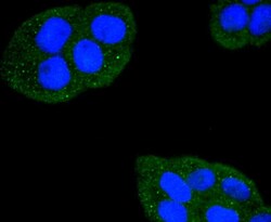
- Immunocytochemical analysis of FAS (CD95) in HepG2 cells using a FAS (CD95) Monoclonal antibody (Product # MA5-32489) as seen in green. The nuclear counter stain is DAPI (blue). Cells were fixed in paraformaldehyde, permeabilised with 0.25% Triton X100/PBS.
- Submitted by
- Invitrogen Antibodies (provider)
- Main image

- Experimental details
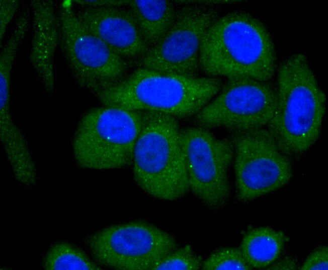
- Immunocytochemical analysis of FAS (CD95) in Hela cells using a FAS (CD95) Monoclonal antibody (Product # MA5-32489) as seen in green. The nuclear counter stain is DAPI (blue). Cells were fixed in paraformaldehyde, permeabilised with 0.25% Triton X100/PBS.

- Submitted by
- Invitrogen Antibodies (provider)
- Main image

- Experimental details
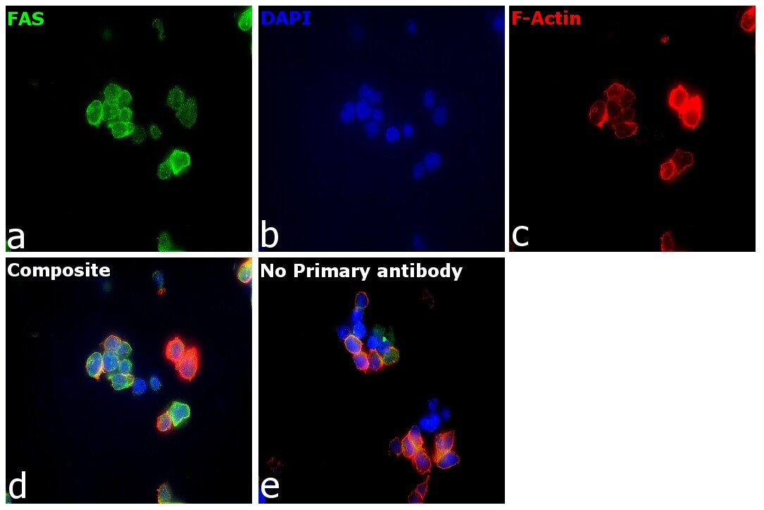
- Immunofluorescence analysis of FAS (Tumor necrosis factor receptor superfamily member 6) was performed using Jurkat cells. The cells were fixed with 4% paraformaldehyde for 10 minutes, permeabilized with 0.1% Triton™ X-100 for 10 minutes, and blocked with 2% BSA for 1 hour at room temperature. The cells were labeled with Fas Recombinant Rabbit Monoclonal Antibody (JJ0942) (Product # MA5-32489) at 1:100 in 0.1% BSA, incubated at 4 degree celsius overnight and then labeled with Donkey anti-Rabbit IgG (H+L) Highly Cross-Adsorbed Secondary Antibody, Alexa Fluor Plus 488 (Product # A32790), (1:2500), for 45 minutes at room temperature (Panel a: Green). Nuclei (Panel b:Blue) were stained with ProLong™ Diamond Antifade Mountant with DAPI (Product # P36962). F-actin (Panel c: Red) was stained with Rhodamine Phalloidin (Product # R415, 1:300). Panel d represents the merged image showing membrane localization. Panel e represents control cells with no primary antibody to assess background. The images were captured at 60X magnification with EVOS™ M7000 Imaging System (Product # AMF7000).
- Submitted by
- Invitrogen Antibodies (provider)
- Main image

- Experimental details
- Immunofluorescence analysis of FAS (Tumor necrosis factor receptor superfamily member 6) was performed using Jurkat cells. The cells were fixed with 4% paraformaldehyde for 10 minutes, permeabilized with 0.1% Triton™ X-100 for 10 minutes, and blocked with 2% BSA for 1 hour at room temperature. The cells were labeled with Fas Recombinant Rabbit Monoclonal Antibody (JJ0942) (Product # MA5-32489) at 1:100 in 0.1% BSA, incubated at 4 degree celsius overnight and then labeled with Donkey anti-Rabbit IgG (H+L) Highly Cross-Adsorbed Secondary Antibody, Alexa Fluor Plus 488 (Product # A32790), (1:2500), for 45 minutes at room temperature (Panel a: Green). Nuclei (Panel b:Blue) were stained with ProLong™ Diamond Antifade Mountant with DAPI (Product # P36962). F-actin (Panel c: Red) was stained with Rhodamine Phalloidin (Product # R415, 1:300). Panel d represents the merged image showing membrane localization. Panel e represents control cells with no primary antibody to assess background. The images were captured at 60X magnification with EVOS™ M7000 Imaging System (Product # AMF7000).

- Submitted by
- Invitrogen Antibodies (provider)
- Main image
- Experimental details
- Immunocytochemical analysis of FAS (CD95) in Hela cells using a FAS (CD95) Monoclonal antibody (Product # MA5-32489) as seen in green. The nuclear counter stain is DAPI (blue). Cells were fixed in paraformaldehyde, permeabilised with 0.25% Triton X100/PBS.

Supportive validation
- Submitted by
- Invitrogen Antibodies (provider)
- Main image

- Experimental details
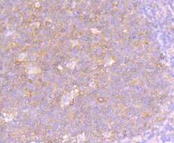
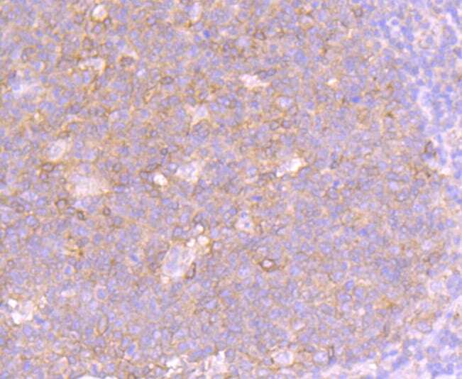
- Immunohistochemical analysis of FAS (CD95) of paraffin-embedded Human tonsil tissue using a FAS-CD95 Monoclonal antibody (Product #MA5-32489). Counter stained with hematoxylin.

- Submitted by
- Invitrogen Antibodies (provider)
- Main image

- Experimental details
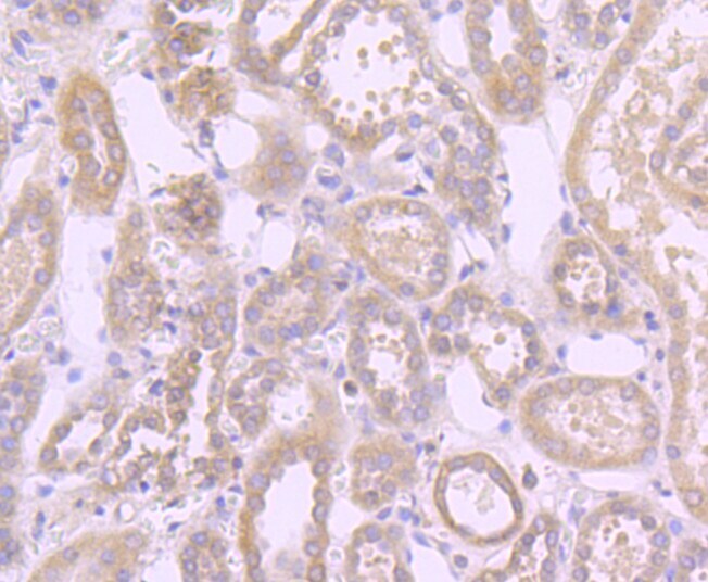
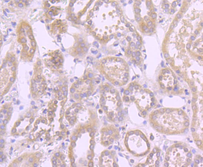
- Immunohistochemical analysis of FAS (CD95) of paraffin-embedded Human liver cancer tissue using a FAS-CD95 Monoclonal antibody (Product #MA5-32489). Counter stained with hematoxylin.
- Submitted by
- Invitrogen Antibodies (provider)
- Main image
- Experimental details
- Immunohistochemical analysis of FAS (CD95) of paraffin-embedded Human tonsil tissue using a FAS-CD95 Monoclonal antibody (Product #MA5-32489). Counter stained with hematoxylin.
- Submitted by
- Invitrogen Antibodies (provider)
- Main image

- Experimental details
- Immunohistochemical analysis of FAS (CD95) of paraffin-embedded Human liver cancer tissue using a FAS-CD95 Monoclonal antibody (Product #MA5-32489). Counter stained with hematoxylin.
Supportive validation
- Submitted by
- Invitrogen Antibodies (provider)
- Main image

- Experimental details
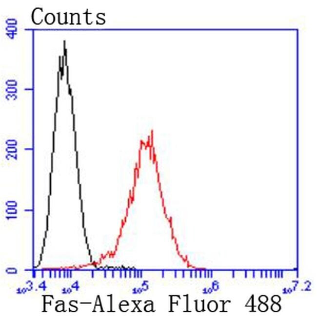
- Flow Cytometric analysis of FAS (CD95) in Raji cells using a FAS (CD95) Monoclonal Antibody (Product # MA5-32489) at a dilution of 1:50, as seen in red compared with an unlabelled control (cells without incubation with primary antibody; black). Alexa Fluor 488-conjugated goat anti rabbit IgG was used as the secondary antibody.
Supportive validation
- Submitted by
- Invitrogen Antibodies (provider)
- Main image
- Experimental details
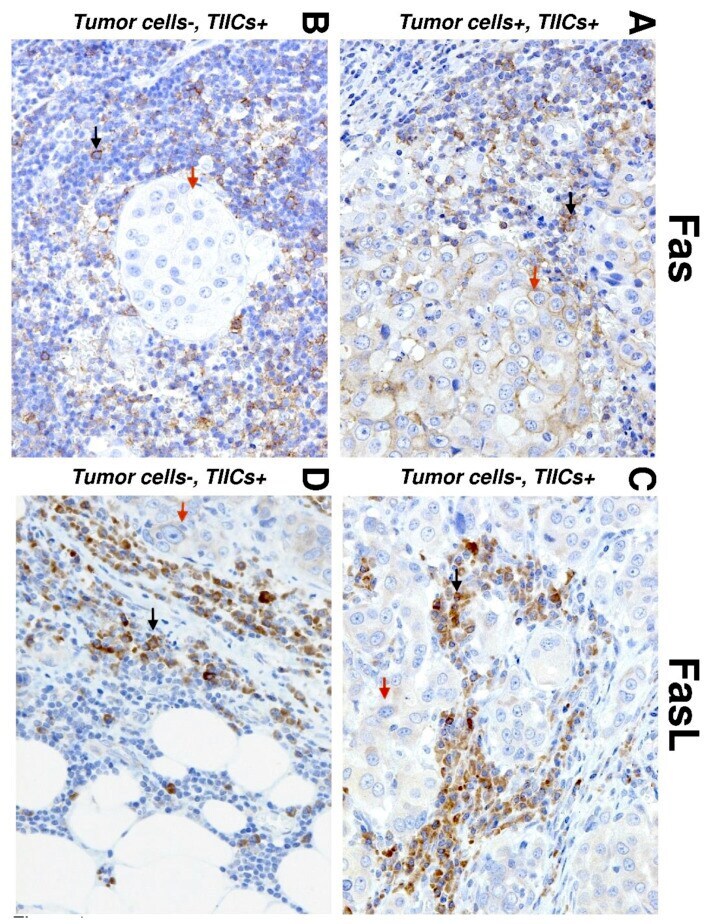
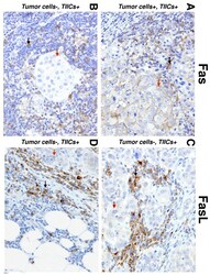
- Figure 1 Immunohistochemistry (IHC) of Fas and Fas ligand (FasL) expression in salivary gland carcinoma (SGC) tissues. Representative images show Fas (left) and FasL (right) staining in tumor cells (red arrows) and tumor-infiltrating immune cells (TIICs) (black arrows) in the tumor periphery. ( A ) IHC shows positive Fas staining in TIICs and tumor cells (20x). ( B ) IHC shows negative Fas staining in tumor cells and positive Fas staining in TIICs (20x). ( C ) IHC shows positive FasL staining in tumor cells and TIICs (20x). ( D ) IHC shows negative FasL staining in tumor cells and positive FasL staining in TIICs (20x).