Explore
 Validate
 Learn
Learn Western blot
Antibody data
- Antibody Data
- Antigen structure
- References [0]
- Comments [0]
- Validations
- Western blot [2]
- Immunocytochemistry [1]
- Immunohistochemistry [2]
- Flow cytometry [1]
Submit
Validation data
Reference
Comment
Report error
- Product number
- AGA-001-200UL - Provider product page

- Provider
- Invitrogen Antibodies
- Product name
- GABA(A) alpha 1 Receptor (extracellular) Polyclonal Antibody
- Antibody type
- Polyclonal
- Antigen
- Other
- Reactivity
- Human, Mouse, Rat
- Host
- Rabbit
- Isotype
- IgG
- Vial size
- 200 µL
- Concentration
- 0.8 mg/mL
- Storage
- -20° C, Avoid Freeze/Thaw Cycles
No comments: Submit comment
Supportive validation
- Submitted by
- Invitrogen Antibodies (provider)
- Main image

- Experimental details
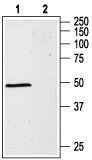
- Western blot analysis of rat brain lysates: - 1. Anti-GABA (A) alpha 1 Receptor (extracellular) Antibody (#AGA-001), (1:1000). 2. Anti-GABA (A) alpha 1 Receptor (extracellular) Antibody , preincubated with GABA (A) alpha 1 Receptor (extracellular) Blocking Peptide (#BLP-GA001).
- Submitted by
- Invitrogen Antibodies (provider)
- Main image

- Experimental details
- Western blot analysis of rat brain lysates: - 1. Anti-GABA (A) alpha 1 Receptor (extracellular) Antibody (#AGA-001), (1:1000). 2. Anti-GABA (A) alpha 1 Receptor (extracellular) Antibody , preincubated with GABA (A) alpha 1 Receptor (extracellular) Blocking Peptide (#BLP-GA001).

Supportive validation
- Submitted by
- Invitrogen Antibodies (provider)
- Main image
- Experimental details
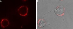
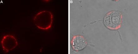
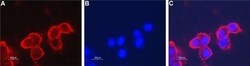
- Expression of GABRA1 in rat PC12 cells - Cell surface detection of GABRA1 in intact living rat pheochromocytoma PC12 cells. A. Extracellular staining of cells using Anti-GABA (A) alpha 1 Receptor (extracellular) Antibody (#AGA-001), (1:100) followed by goat Anti-rabbit-AlexaFluor-594secondary Antibody . B. Merge image of A and live view of the cells.
Supportive validation
- Submitted by
- Invitrogen Antibodies (provider)
- Main image

- Experimental details
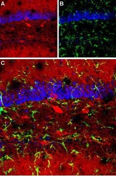
- Expression of GABRA1 in mouse hippocampus - Immunohistochemical staining of mouse hippocampus using Anti-GABA (A) alpha 1 Receptor (extracellular) Antibody (#AGA-001). A. Distribution of GABRA1 (red). B. Distribution of glial fibrillary acidic protein (green). C. Merge of the two images indicates that distribution of GABRA1 is restricted to neurons and their processes. DAPI is used as the counterstain (blue).
- Submitted by
- Invitrogen Antibodies (provider)
- Main image
- Experimental details
- Colocalization of CaV1.2 and GABA (A) alpha 1 Receptor in rat cerebellum - Immunohistochemical staining of rat cerebellum using Guinea pig Anti-CaV1.2 (CACNA1C) Antibody (#ACC-003-GP) and Anti-GABA (A) alpha 1 Receptor (extracellular) Antibody (#AGA-001). A. CaV1.2 (green) is detected in the granule layer of the cerebellum (G) and in the upper molecular layer (star). B. In the same section,GABRA1 (red) is seen in the granule layer. C. Merge of the two images reveals high degree of colocalization between CaV1.2 and GABRA1 in the granule layer.

Supportive validation
- Submitted by
- Invitrogen Antibodies (provider)
- Main image

- Experimental details
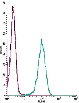
- Cell surface detection ofGABRA1by indirect flow cytometry in live intact human THP-1monocyticleukemia cells: - (black line) cells. (red) Cells+ goat- Anti-rabbit-FITC. (green) Cells + Anti-GABA (A) alpha 1 Receptor (extracellular) Antibody (#AGA-001), (2.5μg) + goat- Anti-rabbit-FITC.