 Explore
 Validate
 Learn
Learn Western blot
 ELISA
ELISAAntibody data
- Antibody Data
- Antigen structure
- References [0]
- Comments [0]
- Validations
- Western blot [3]
- Immunocytochemistry [1]
Submit
Validation data
Reference
Comment
Report error
- Product number
- PA1-32487 - Provider product page

- Provider
- Invitrogen Antibodies
- Product name
- BMI-1 Polyclonal Antibody
- Antibody type
- Polyclonal
- Antigen
- Synthetic peptide
- Description
- Store product as a concentrated solution. Centrifuge briefly prior to opening the vial.
- Reactivity
- Human
- Host
- Goat
- Isotype
- IgG
- Vial size
- 50 µg
- Concentration
- 0.91 mg/mL
- Storage
- Store at 4°C short term. For long term storage, store at -20°C, avoiding freeze/thaw cycles.
No comments: Submit comment
Supportive validation
- Submitted by
- Invitrogen Antibodies (provider)
- Main image

- Experimental details
- Western blot detection of BMI-1 in 20 µg of U2OS whole cell lysate (bone osteosarcoma). Sample was separated by 4-20% SDS-PAGE and transferred onto nitrocellulose. After blocking in PBS containing 5% nonfat dry milk, the membrane was probed using a BMI-1 polyclonal antibody (Product # PA1-32487) at a dilution of 1:1000 in PBS containing 1% nonfat dry milk after incubation overnight at 4ºC. The membrane was washed and reacted with a 1:20,000 dilution of IR Dye800 conjugated rabbit anti-Goat IgG (H&L) for 45 min at room temperature.

- Submitted by
- Invitrogen Antibodies (provider)
- Main image

- Experimental details
- Western blot using BMI-1 Polyclonal Antibody (Product # PA1-32487) shows detection of a band at 37 kDa corresponding to human Bmi1 (arrowhead). Approximately 20 µg of a U2OS whole cell lysate was separated by 4-20% SDS-PAGE and transferred onto nitrocellulose. After blocking in PBS containing 5% nonfat dry milk, the membrane was probed overnight at 4° C with the primary antibody diluted to 1:1,000 in PBS containing 1% nonfat dry milk. The membrane was washed and reacted with a 1:20,000 dilution of a rabbit anti-Goat IgG [H&L] for 45 min at room temperature.

- Submitted by
- Invitrogen Antibodies (provider)
- Main image
- Experimental details
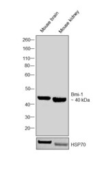
- Western blot was performed using Anti-BMI-1 Polyclonal Antibody (Product # PA1-32487) and a 40 kDa band corresponding to BMI-1 was observed across tissues tested. Whole Cell Extract (40 µg lysate) of Mouse Brain (Lane 1), Mouse Kidney (Lane 2) were electrophoresed using NuPAGE™ 4-12% Bis-Tris Protein Gel (Product # NP0321BOX). Resolved proteins were then transferred onto a Nitrocellulose membrane (Product # IB23001) by iBlot® 2 Dry Blotting System (Product # IB21001). The blot was probed with the primary antibody (1:1000 dilution) and detected by chemiluminescence with Rabbit anti-Goat IgG (H+L) Superclonal™ Recombinant Secondary Antibody, HRP (Product # A27014, 1:4000 dilution) using the iBright FL 1000 (Product # A32752). Chemiluminescent detection was performed using Novex® ECL Chemiluminescent Substrate Reagent Kit (Product # WP20005).

Supportive validation
- Submitted by
- Invitrogen Antibodies (provider)
- Main image

- Experimental details
- Immunocytochemistry-Immunofluorescence analysis of BMI-1 in methanol fixed (100%, 5 min) HepG2 cells. The cells were blocked and permeabilized in 1%BSA / 10% normal donkey serum / 0.3M glycine in 0.1% PBS-Tween for 1h prior to incubation with the BMI-1 Polyclonal Antibody (Product # PA1-32487) (1:200 dilution) overnight at 4°C and detected with a 488nm fluorescent dye conjugated secondary Ab. Cell nuclei are stained with DAPI (blue) and plasma membranes are stained with WGA (red).
