Explore
 Validate
 Learn
Learn Immunohistochemistry
Antibody data
- Antibody Data
- Antigen structure
- References [2]
- Comments [0]
- Validations
- Immunohistochemistry [2]
Submit
Validation data
Reference
Comment
Report error
- Product number
- MAB33342 - Provider product page

- Provider
- Novus Biologicals
- Product name
- Mouse Monoclonal BMI-1 Antibody
- Antibody type
- Monoclonal
- Description
- Protein A or G purified from hybridoma culture supernatant. Detects human BMI-1 in direct ELISAs.
- Reactivity
- Human
- Host
- Mouse
- Isotype
- IgG
- Vial size
- 100 ug
- Concentration
- LYOPH
- Storage
- Use a manual defrost freezer and avoid repeated freeze-thaw cycles. 12 months from date of receipt, -20 to -70 degreesC as supplied. 1 month, 2 to 8 degreesC under sterile conditions after reconstitution. 6 months, -20 to -70 degreesC under sterile conditions after reconstitution.
Submitted references FUN14 domain-containing 1 promotes breast cancer proliferation and migration by activating calcium-NFATC1-BMI1 axis.
p53 regulates epithelial-mesenchymal transition and stem cell properties through modulating miRNAs.
Wu L, Zhang D, Zhou L, Pei Y, Zhuang Y, Cui W, Chen J
EBioMedicine 2019 Mar;41:384-394
EBioMedicine 2019 Mar;41:384-394
p53 regulates epithelial-mesenchymal transition and stem cell properties through modulating miRNAs.
Chang CJ, Chao CH, Xia W, Yang JY, Xiong Y, Li CW, Yu WH, Rehman SK, Hsu JL, Lee HH, Liu M, Chen CT, Yu D, Hung MC
Nature cell biology 2011 Mar;13(3):317-23
Nature cell biology 2011 Mar;13(3):317-23
No comments: Submit comment
Supportive validation
- Submitted by
- Novus Biologicals (provider)
- Main image

- Experimental details
- BMI-1 in Human Breast. BMI-1 was detected in immersion fixed paraffin-embedded sections of normal human breast using Human BMI-1 Monoclonal Antibody (Catalog # MAB33342) at 15 µg/mL overnight at 4 °C. Tissue was stained using the Anti-Mouse HRP-DAB Cell & Tissue Staining Kit (brown; Catalog # CTS002) and counterstained with hematoxylin (blue). Lower panel shows a lack of labeling if primary antibodies are omitted and tissue is stained only with secondary antibody followed by incubation with detection reagents. View our protocol for Chromogenic IHC Staining of Paraffin-embedded Tissue Sections.

- Submitted by
- Novus Biologicals (provider)
- Main image
- Experimental details
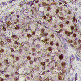
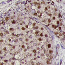
- BMI-1 in Human Breast Cancer Tissue. BMI-1 was detected in immersion fixed paraffin-embedded sections of human breast cancer tissue using Human BMI-1 Monoclonal Antibody (Catalog # MAB33342) at 25 µg/mL overnight at 4 °C. Tissue was stained using the Anti-Mouse HRP-DAB Cell & Tissue Staining Kit (brown; Catalog # CTS002) and counterstained with hematoxylin (blue). Specific labeling was localized to the nuclei of epithelial cells. View our protocol for Chromogenic IHC Staining of Paraffin-embedded Tissue Sections.